File:Lymph node histology 05.jpg: Difference between revisions
From Embryology
| Line 8: | Line 8: | ||
* "high" from the thickness (low cuboidal) of the endothelial cells. | * "high" from the thickness (low cuboidal) of the endothelial cells. | ||
** endothelial cells are typically a squamous epithelium. | ** endothelial cells are typically a squamous epithelium. | ||
* vascular site for lymphocyte entry | * vascular site for lymphocyte entry and exit from secondary lymphoid organs. | ||
* endothelial cells express ligands that bind lymphocytes, aiding their adhesion and subsequent transmigration (lymphocyte extravasation) into the lymph node. | |||
Revision as of 17:41, 2 March 2014
Lymph Node Histology - High Endothelial Venules
(Stain - Haematoxylin Eosin)
High Endothelial Venules (HEV)
- specialized postcapillary venules.
- "high" from the thickness (low cuboidal) of the endothelial cells.
- endothelial cells are typically a squamous epithelium.
- vascular site for lymphocyte entry and exit from secondary lymphoid organs.
- endothelial cells express ligands that bind lymphocytes, aiding their adhesion and subsequent transmigration (lymphocyte extravasation) into the lymph node.
- Lymph Node Histology: Subcapsular Sinus | Follicle | Germinal Centre | Medullary Cords and Sinuses | High Endothelial Venules | Macrophages | Node cartoons
Links: Histology | Histology Stains | Blue Histology images copyright Lutz Slomianka 1998-2009. The literary and artistic works on the original Blue Histology website may be reproduced, adapted, published and distributed for non-commercial purposes. See also the page Histology Stains.
Cite this page: Hill, M.A. (2024, June 24) Embryology Lymph node histology 05.jpg. Retrieved from https://embryology.med.unsw.edu.au/embryology/index.php/File:Lymph_node_histology_05.jpg
- © Dr Mark Hill 2024, UNSW Embryology ISBN: 978 0 7334 2609 4 - UNSW CRICOS Provider Code No. 00098G
File history
Yi efo/eka'e gwa ebo wo le nyangagi wuncin ye kamina wunga tinya nan
| Gwalagizhi | Nyangagi | Dimensions | User | Comment | |
|---|---|---|---|---|---|
| current | 18:30, 25 February 2012 |  | 450 × 600 (87 KB) | Z8600021 (talk | contribs) | increase image size |
| 09:03, 14 February 2011 | 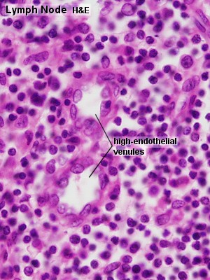 | 300 × 400 (52 KB) | S8600021 (talk | contribs) | ==Lymph Node Histology== Original file name: Lyn42he.jpg http://www.lab.anhb.uwa.edu.au/mb140/CorePages/Lymphoid1/lymph1.htm#Lymph Lymph node histology 05.jpg {{Template:Blue Histology}} Category:Immune |
You cannot overwrite this file.