File:Sabin1909 fig17.jpg: Difference between revisions
From Embryology
(==Fig 17. and Fig 2. Human embryo measuring 50 mm== Fig. 17. Sagittal section of a human embyo measuring 50 mm., Mall collection, No. 96, showing the posterior lymph sac within the pelvis and its extension along the femoral vein. X about 8. F., femur; Lg) |
mNo edit summary |
||
| Line 1: | Line 1: | ||
==Fig 17. and Fig 2. Human embryo measuring 50 mm== | ==Fig 17. and Fig 2. Human embryo measuring 50 mm== | ||
Fig. 17. Sagittal section of a human embyo measuring 50 mm | Fig. 17. Sagittal section of a human embyo measuring 50 mm. | ||
Mall collection, No. 96, showing the posterior lymph sac within the pelvis and its extension along the femoral vein. X about 8. F., femur; Lg., lymphoglandnla (femoralis) ; 0. s., OS sacrum ; S. l. p., saccus lymphaticus posterior with lymph node in the border; V. a., vena sciatica; V. l. v., vertebra lumbalis v. | |||
==Reference== | |||
===Reference=== | |||
Florence R. Sabin, The lymphatic system in human embryos, with a consideration of the morphology of the system as a whole. American Journal of Anatomy Volume 9, Issue 1, pages 43–91, 1909 | Florence R. Sabin, The lymphatic system in human embryos, with a consideration of the morphology of the system as a whole. American Journal of Anatomy Volume 9, Issue 1, pages 43–91, 1909 | ||
[[Category:Historic Embryology]] [[Category:Human]] [[Category:Immune]] [[Category:Cardiovascular]] | [[Category:Historic Embryology]] [[Category:Human]] [[Category:Immune]] [[Category:Cardiovascular]] | ||
Latest revision as of 12:41, 27 January 2014
Fig 17. and Fig 2. Human embryo measuring 50 mm
Fig. 17. Sagittal section of a human embyo measuring 50 mm.
Mall collection, No. 96, showing the posterior lymph sac within the pelvis and its extension along the femoral vein. X about 8. F., femur; Lg., lymphoglandnla (femoralis) ; 0. s., OS sacrum ; S. l. p., saccus lymphaticus posterior with lymph node in the border; V. a., vena sciatica; V. l. v., vertebra lumbalis v.
Reference
Florence R. Sabin, The lymphatic system in human embryos, with a consideration of the morphology of the system as a whole. American Journal of Anatomy Volume 9, Issue 1, pages 43–91, 1909
File history
Click on a date/time to view the file as it appeared at that time.
| Date/Time | Thumbnail | Dimensions | User | Comment | |
|---|---|---|---|---|---|
| current | 14:06, 30 March 2011 | 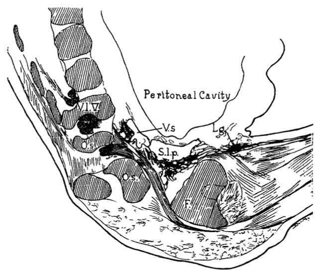 | 645 × 545 (80 KB) | S8600021 (talk | contribs) | ==Fig 17. and Fig 2. Human embryo measuring 50 mm== Fig. 17. Sagittal section of a human embyo measuring 50 mm., Mall collection, No. 96, showing the posterior lymph sac within the pelvis and its extension along the femoral vein. X about 8. F., femur; Lg |
You cannot overwrite this file.
File usage
There are no pages that use this file.