File:Pancreas acinar cell em01.jpg: Difference between revisions
From Embryology
No edit summary |
|||
| Line 5: | Line 5: | ||
:'''Links:''' [[:File:Pancreas_histology_002.jpg|Acinar cell - light microscope]] | [[:File:Pancreas_acinar_cell_em01.jpg|Acinar cell - electron microscope]] | | :'''Links:''' [[:File:Pancreas_histology_002.jpg|Acinar cell - light microscope]] | [[:File:Pancreas_acinar_cell_em01.jpg|Acinar cell - electron microscope]] | [[Gastrointestinal Tract - Pancreas Histology|Pancreas Histology]] | [[Gastrointestinal Tract - Pancreas Development|Pancreas Development]] | ||
===Reference=== | ===Reference=== | ||
Revision as of 15:34, 25 February 2013
Pancreas Acinar Cell
This cell has been labeled to show cell structures in the cytoplasm and nucleus.
Acinar cells are part of the digestive exocrine function of the pancreas. The cytoplasm of these cells has extensive rough endoplasmic reticulum (for protein synthesis) and contain many zymogen granules (for inactive enzyme storage).
- Links: Acinar cell - light microscope | Acinar cell - electron microscope | Pancreas Histology | Pancreas Development
Reference
Dartmouth Images 18_PancreasMito20kX_92.jpg
File history
Yi efo/eka'e gwa ebo wo le nyangagi wuncin ye kamina wunga tinya nan
| Gwalagizhi | Nyangagi | Dimensions | User | Comment | |
|---|---|---|---|---|---|
| current | 15:26, 25 February 2013 | 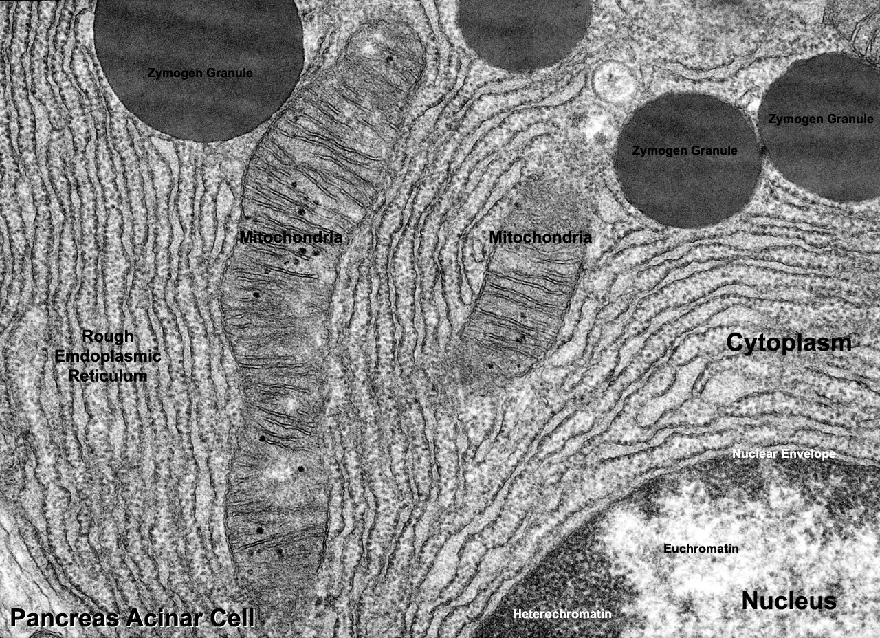 | 1,280 × 928 (496 KB) | Z8600021 (talk | contribs) | ==Pancreas Acinar Cell== This cell has been labeled to show cell structures in the cytoplasm and nucleus. Acinar cells are part of the digestive exocrine function of the pancreas. The cytoplasm of these cells has extensive rough endoplasmic reticulum (fo |
You cannot overwrite this file.
File usage
The following page uses this file: