File:Lymph node histology 01.jpg: Difference between revisions
From Embryology
No edit summary |
No edit summary |
||
| (3 intermediate revisions by the same user not shown) | |||
| Line 1: | Line 1: | ||
==Lymph Node Histology== | ==Lymph Node Histology== | ||
{{HE}} | |||
* '''Reticular cells''' (and reticular fibres) form a delicate network between the capsule and trabeculae. | |||
* Only their large and light nuclei are easily visible in the microscope. | |||
* The cytoplasm of reticular cells is only weakly eosinophilic. | |||
* Lymphocytes and macrophages are housed in the network of reticular cells and the reticular fibres formed by them. | |||
* The processes of reticular cells and reticular fibres extend into and criss-cross within the sinuses. | |||
Latest revision as of 15:34, 24 February 2013
Lymph Node Histology
(Stain - Haematoxylin Eosin)
- Reticular cells (and reticular fibres) form a delicate network between the capsule and trabeculae.
- Only their large and light nuclei are easily visible in the microscope.
- The cytoplasm of reticular cells is only weakly eosinophilic.
- Lymphocytes and macrophages are housed in the network of reticular cells and the reticular fibres formed by them.
- The processes of reticular cells and reticular fibres extend into and criss-cross within the sinuses.
- Lymph Node Histology: Subcapsular Sinus | Follicle | Germinal Centre | Medullary Cords and Sinuses | High Endothelial Venules | Macrophages | Node cartoons
Links: Histology | Histology Stains | Blue Histology images copyright Lutz Slomianka 1998-2009. The literary and artistic works on the original Blue Histology website may be reproduced, adapted, published and distributed for non-commercial purposes. See also the page Histology Stains.
Cite this page: Hill, M.A. (2024, June 21) Embryology Lymph node histology 01.jpg. Retrieved from https://embryology.med.unsw.edu.au/embryology/index.php/File:Lymph_node_histology_01.jpg
- © Dr Mark Hill 2024, UNSW Embryology ISBN: 978 0 7334 2609 4 - UNSW CRICOS Provider Code No. 00098G
File history
Yi efo/eka'e gwa ebo wo le nyangagi wuncin ye kamina wunga tinya nan
| Gwalagizhi | Nyangagi | Dimensions | User | Comment | |
|---|---|---|---|---|---|
| current | 18:32, 25 February 2012 | 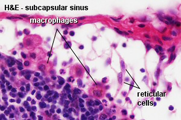 | 600 × 400 (61 KB) | Z8600021 (talk | contribs) | increase image size |
| 08:57, 14 February 2011 |  | 300 × 200 (25 KB) | S8600021 (talk | contribs) | ==Lymph Node Histology== :"Reticular cells (and reticular fibres) form a delicate network between the capsule and trabeculae. Only their large and light nuclei are easily visible in the microscope. The cytoplasm of reticular cells is only weakly eosinoph |
You cannot overwrite this file.