File:Gray0653.jpg: Difference between revisions
No edit summary |
|||
| Line 2: | Line 2: | ||
From model by His. | From model by His. | ||
The corpus striatum (Figs. 651 and 653) appears in the fourth week as a triangular thickening of the floor of the telencephalon between the optic recess and the interventricular foramen, and continuous behind with the thalamic part of the diencephalon. It increases in size, and by the second month is seen as a swelling in the floor of the future lateral ventricle; this swelling reaches as far as the posterior end of the primitive hemisphere, and when this part of the hemisphere grows backward and downward to form the temporal lobe, the posterior part of the corpus striatum is carried into the roof of the inferior horn of the ventricle, where it is seen as the tail of the caudate nucleus in the adult brain. During the fourth and fifth months the corpus striatum becomes incompletely subdivided by the fibers of the internal capsule into two masses, an inner, the caudate nucleus, and an outer, the lentiform nucleus. In front, the corpus striatum is continuous with the anterior perforated substance; laterally it is confluent for a time with that portion of the wall of the vesicle which is developed into the insula, but this continuity is subsequently interrupted by the fibers of the external capsule. | |||
Revision as of 10:44, 15 February 2013
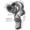
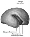
Interior of brain of human embryo of five weeks
From model by His.
The corpus striatum (Figs. 651 and 653) appears in the fourth week as a triangular thickening of the floor of the telencephalon between the optic recess and the interventricular foramen, and continuous behind with the thalamic part of the diencephalon. It increases in size, and by the second month is seen as a swelling in the floor of the future lateral ventricle; this swelling reaches as far as the posterior end of the primitive hemisphere, and when this part of the hemisphere grows backward and downward to form the temporal lobe, the posterior part of the corpus striatum is carried into the roof of the inferior horn of the ventricle, where it is seen as the tail of the caudate nucleus in the adult brain. During the fourth and fifth months the corpus striatum becomes incompletely subdivided by the fibers of the internal capsule into two masses, an inner, the caudate nucleus, and an outer, the lentiform nucleus. In front, the corpus striatum is continuous with the anterior perforated substance; laterally it is confluent for a time with that portion of the wall of the vesicle which is developed into the insula, but this continuity is subsequently interrupted by the fibers of the external capsule.
- Brain Development Links: Week 4.5 exterior | Week 5 exterior | Week 5 interior | 3 month | 3 month hindbrain | 4 month | 5 month | Gray's Neural Images | Neural System Development
651 Human Embryo Brain (week 4.5 exterior view)
652 Human Embryo Brain (week 5 exterior view)
653 Human Embryo Brain (week 5 interior view)

654 Human Fetal Brain (3 months)
655 Human Fetal Brain (4 months)
658 Human Fetal Brain (5 months)


- Gray's Images: Development | Lymphatic | Neural | Vision | Hearing | Somatosensory | Integumentary | Respiratory | Gastrointestinal | Urogenital | Endocrine | Surface Anatomy | iBook | Historic Disclaimer
| Historic Disclaimer - information about historic embryology pages |
|---|
 |
| iBook - Gray's Embryology | |
|---|---|

|
|
Reference
Gray H. Anatomy of the human body. (1918) Philadelphia: Lea & Febiger.
Cite this page: Hill, M.A. (2024, June 26) Embryology Gray0653.jpg. Retrieved from https://embryology.med.unsw.edu.au/embryology/index.php/File:Gray0653.jpg
- © Dr Mark Hill 2024, UNSW Embryology ISBN: 978 0 7334 2609 4 - UNSW CRICOS Provider Code No. 00098G
File history
Yi efo/eka'e gwa ebo wo le nyangagi wuncin ye kamina wunga tinya nan
| Gwalagizhi | Nyangagi | Dimensions | User | Comment | |
|---|---|---|---|---|---|
| current | 08:00, 20 May 2012 |  | 698 × 700 (108 KB) | Z8600021 (talk | contribs) | ==Interior of brain of human embryo of five weeks== From model by His. {{Gray Anatomy}} Category:Human Category:Historic Embryology Category:Week 5 Category:Gray's 1918 Anatomy Category:Neural |
You cannot overwrite this file.
File usage
The following 14 pages use this file:
- Anatomy of the Human Body by Henry Gray
- BGD Lecture - Endocrine Development
- Lecture - Endocrine Development
- REI - Reproductive Medicine Seminar 2018
- Royal Hospital for Women - Reproductive Medicine Seminar 2018
- User:Z5019799
- File:Gray0649.jpg
- File:Gray0651.jpg
- File:Gray0652.jpg
- File:Gray0653.jpg
- File:Gray0654.jpg
- File:Gray0655.jpg
- File:Gray0658.jpg
- Template:Gray-Brain