File:Buell01.jpg: Difference between revisions
From Embryology
No edit summary |
No edit summary |
||
| (One intermediate revision by the same user not shown) | |||
| Line 1: | Line 1: | ||
==Fig. 1. Median-sagittal section | ==Fig. 1. Median-sagittal section through tip of lung-bud of a 20 somite chick== | ||
Median-sagittal section (10 micron in thickness) through tip of lung-bud of a 20-somite chick, 40 hours' | Median-sagittal section (10 micron in thickness) through tip of lung-bud of a 20-somite chick, 40 hours' incubation haematoxylin and eosin stain, series B. | ||
Angioblasts, forerunners of pulmonary system, are seen proliferating from and near dorsal endothelial wall of sinus venosus. They extend dorsally toward the ventral surface of the gut, which shows a slight ventral swelling - the primary lung rudiment. This is about the earliest stage in which there is any evidence of the formation of a pulmonary vascular system. | Angioblasts, forerunners of pulmonary system, are seen proliferating from and near dorsal endothelial wall of sinus venosus. They extend dorsally toward the ventral surface of the gut, which shows a slight ventral swelling - the primary lung rudiment. This is about the earliest stage in which there is any evidence of the formation of a pulmonary vascular system. | ||
Latest revision as of 08:42, 24 Ocak 2013
Fig. 1. Median-sagittal section through tip of lung-bud of a 20 somite chick
Median-sagittal section (10 micron in thickness) through tip of lung-bud of a 20-somite chick, 40 hours' incubation haematoxylin and eosin stain, series B.
Angioblasts, forerunners of pulmonary system, are seen proliferating from and near dorsal endothelial wall of sinus venosus. They extend dorsally toward the ventral surface of the gut, which shows a slight ventral swelling - the primary lung rudiment. This is about the earliest stage in which there is any evidence of the formation of a pulmonary vascular system.
- 1922 Chicken Pulmonary: Fig 1 | Fig 2 | Fig 3 | Fig 4 | Fig 5 | Fig 6 | Fig 7 | Fig 8 | Fig 9 | Fig 10 | Plate 1 | Plate 2 | Carnegie No.66 | Chicken Development | Respiratory
- Links: Carnegie Institution of Washington - Contributions to Embryology | Chicken Development | Respiratory System Development
| Historic Disclaimer - information about historic embryology pages |
|---|
 |
File history
Yi efo/eka'e gwa ebo wo le nyangagi wuncin ye kamina wunga tinya nan
| Gwalagizhi | Nyangagi | Dimensions | User | Comment | |
|---|---|---|---|---|---|
| current | 19:12, 23 Ocak 2013 | 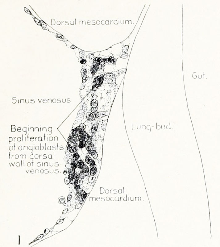 | 713 × 800 (70 KB) | Z8600021 (talk | contribs) | {{Buell1922}} :'''Links:''' Carnegie Institution of Washington - Contributions to Embryology | Chicken Development | Respiratory System Development {{Historic Disclaimer}} Category:Cardiovascular [[ |
You cannot overwrite this file.
File usage
The following page uses this file:


