File:Chicken primordial germ cell migration model.jpg: Difference between revisions
(==Chicken Primordial Germ Cell (PGC) Migration Model== At HH12–13, the yolk sac circulation courses in loop (red arrows) to enter the embryo via the heart. At this stage, the majority of PGCs (green dots) localized axially at the border between the are) |
|||
| (One intermediate revision by the same user not shown) | |||
| Line 1: | Line 1: | ||
==Chicken Primordial Germ Cell (PGC) Migration Model== | ==Chicken Primordial Germ Cell (PGC) Migration Model== | ||
'''HH12–13''' - yolk sac circulation courses in loop (red arrows) to enter the embryo via the heart. The majority of PGCs (green dots) localized axially at the border between the area opaca and pellucida, where the sinus terminalis converged in the anterior vitelline veins. | |||
'''HH14–16''' - PGCs (green dots) circulated effectively towards the embryo via the sinus terminalis and the anterior vitelline veins towards the heart. Thereafter, the PGCs traffic via the aorta to the caudal part of the embryo and become lodged in the genital ridges. | |||
HH - Hamburger Hamilton Stage | |||
:'''Links:''' [[Chicken Development]] | [[Genital System Development|Genital Development]] | |||
===Reference=== | ===Reference=== | ||
<pubmed>23213395</pubmed>| [http://www.ncbi.nlm.nih.gov/pmc/articles/PMC3507194 PMC3507194] | [http://bio.biologists.org/content/1/11/1146 Biol Open] | |||
====Copyright==== | |||
This is an Open Access article distributed under the terms of the Creative Commons Attribution Non-Commercial Share Alike License (http://creativecommons.org/licenses/by-nc-sa/3.0/). | |||
http://bio.biologists.org/content/1/11/1146/F5.expansion.html (panel F cropped from full figure) | |||
[[Category:Chicken]] [[Category:Genital]] | |||
Latest revision as of 09:03, 22 December 2012
Chicken Primordial Germ Cell (PGC) Migration Model
HH12–13 - yolk sac circulation courses in loop (red arrows) to enter the embryo via the heart. The majority of PGCs (green dots) localized axially at the border between the area opaca and pellucida, where the sinus terminalis converged in the anterior vitelline veins.
HH14–16 - PGCs (green dots) circulated effectively towards the embryo via the sinus terminalis and the anterior vitelline veins towards the heart. Thereafter, the PGCs traffic via the aorta to the caudal part of the embryo and become lodged in the genital ridges.
HH - Hamburger Hamilton Stage
- Links: Chicken Development | Genital Development
Reference
<pubmed>23213395</pubmed>| PMC3507194 | Biol Open
Copyright
This is an Open Access article distributed under the terms of the Creative Commons Attribution Non-Commercial Share Alike License (http://creativecommons.org/licenses/by-nc-sa/3.0/).
http://bio.biologists.org/content/1/11/1146/F5.expansion.html (panel F cropped from full figure)
File history
Yi efo/eka'e gwa ebo wo le nyangagi wuncin ye kamina wunga tinya nan
| Gwalagizhi | Nyangagi | Dimensions | User | Comment | |
|---|---|---|---|---|---|
| current | 08:58, 22 December 2012 | 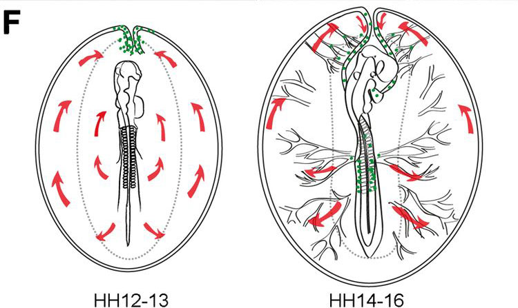 | 750 × 447 (69 KB) | Z8600021 (talk | contribs) | ==Chicken Primordial Germ Cell (PGC) Migration Model== At HH12–13, the yolk sac circulation courses in loop (red arrows) to enter the embryo via the heart. At this stage, the majority of PGCs (green dots) localized axially at the border between the are |
You cannot overwrite this file.
File usage
The following page uses this file: