Urogenital Sinus Movie

From Embryology
Embryology - 19 Mar 2024    Facebook link Pinterest link Twitter link  Expand to Translate  
Google Translate - select your language from the list shown below (this will open a new external page)

العربية | català | 中文 | 中國傳統的 | français | Deutsche | עִברִית | हिंदी | bahasa Indonesia | italiano | 日本語 | 한국어 | မြန်မာ | Pilipino | Polskie | português | ਪੰਜਾਬੀ ਦੇ | Română | русский | Español | Swahili | Svensk | ไทย | Türkçe | اردو | ייִדיש | Tiếng Việt    These external translations are automated and may not be accurate. (More? About Translations)

<html5media height="520" width="360">File:Urogenital_sinus_001.mp4</html5media>

Click Here to play on mobile device

Urogenital sinus 001 icon.jpg
Urogenital Sinus and Renal Development

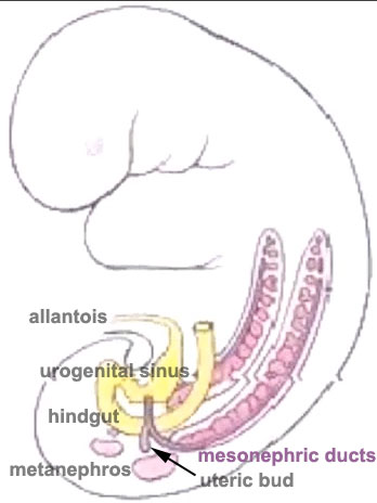
This animation gives an overview of both early renal and genital (urogenital) development associated with the urogenital sinus.

First observe the development of the intermediate mesoderm.

  • This animation shows the 3 stages of kidney development (pink) in relation to the hindgut region between Week 4 and 5 (GA 6 and 7).
  • earliest stage of kidney development begins up near the pharyngeal arches as the pronephros (cervical nephrotomes) which quickly degenerate, beside these the mesonephric duct begins to form (purple).
  • middle stage is the extensive mesonephros (red) which extends down towards the developing hindgut region (yellow). Associated with the mesonephros is the mesonephric duct (purple).
  • The mesonephric duct gives off a lateral branch forming the ureteric bud.
  • final stage the ureteric bud interacts with the surrounding mesenchyme to begin the final stage in renal development, formation of the metanephros (pink) which will form the adult kidney.


Next observe the changes occuring in the hindgut endoderm region (yellow) ending in the embryo at the cloaca.

  • The posterior portion (nearest the mesonephros) remains as the gastrointestinal tract hindgut forming the rectum.
  • The anterior portion (extends into the connecting stalk as the allantois)
    • becomes separated from the hindgut and forms the urogenital sinus, primordia of the urinary bladder.
    • The two mesonephric ducts initially open into the urogenital sinus.


Links: MP4 movie | Renal Movies | Genital Movies | Renal System Development
Urogenital sinus 002.jpg Urogenital sinus 003.jpg


Glossary Links: A | B | C | D | E | F | G | H | I | J | K | L | M | N | O | P | Q | R | S | T | U | V | W | X | Y | Z | Numbers | Symbols | Movies


Cite this page: Hill, M.A. (2024, March 19) Embryology Urogenital Sinus Movie. Retrieved from https://embryology.med.unsw.edu.au/embryology/index.php/Urogenital_Sinus_Movie

What Links Here?
© Dr Mark Hill 2024, UNSW Embryology ISBN: 978 0 7334 2609 4 - UNSW CRICOS Provider Code No. 00098G