Skeletal System 3D stage 22 Movie
| Embryology - 28 Apr 2024 |
|---|
| Google Translate - select your language from the list shown below (this will open a new external page) |
|
العربية | català | 中文 | 中國傳統的 | français | Deutsche | עִברִית | हिंदी | bahasa Indonesia | italiano | 日本語 | 한국어 | မြန်မာ | Pilipino | Polskie | português | ਪੰਜਾਬੀ ਦੇ | Română | русский | Español | Swahili | Svensk | ไทย | Türkçe | اردو | ייִדיש | Tiếng Việt These external translations are automated and may not be accurate. (More? About Translations) |
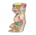
| <html5media height="700" width="700">File:Stage22_SKM3d.mp4</html5media> |
The skeletal system is primarily mesodermal in origin, with additional input from the neural crest. The neural crest contributes to the craniofacial skeleton, with somites and the lateral plate mesoderm forming the axial skeleton and the limbs respectively. Below the animation is a more complete description of each system. Colour code:
|
Skeletal System Development
The skeletal system is primarily mesodermal in origin, with additional input from the neural crest. The neural crest contributes to the craniofacial skeleton, with somites and the lateral plate mesoderm forming the axial skeleton and the limbs respectively.
The first stage in bone development is migration of preskeletal cells to the correct anatomical location. This is associated with an epithelium and basement membrane, which interact with the preskeletal cells, causing them to undergo condensation. Cell condensation is the process whereby populations of cells gather together and differentiate into a single cell type, be it cartilage, bone, muscle, tendon etc... Adhesion molecules, epithelial cell surface receptors and extracellular matrix molecules initiate condensation and set its boundaries. The next step in bone formation is differentiation of the cells to chondrocytes or osteoblasts.
2 types of Ossification can occur:
Intramembranous – primarily in the skull Endochondrial – the majority of bones, particularly the vertebrae, limb long bones etc… The difference between the two is that endochondrial ossification involves formation of a cartilage template which later ossifies to bone, while intramembranous ossification bypasses this step, with the ossification taking place over a membranous template.
The cartilage template formed in endochondrial ossification by chondrocytes. Unlike bone, cartilage is avascular, thus an important step in ossification is development of vasculature. This achieved by the chondrocytes which hypertrophy and begin to secrete Vascular Endothelial Growth Factor (VEGF), subsequently undergoing apoptosis. VEGF induces vasculature to form, bringing with it immature osteoblasts, which begin to form bone.
|
|
|
|
|
|
These 3d movies were part of the UNSW Medical degree Independent Learning Project (ILP) prepared by Aashish Kumar (2006).
Glossary Links: A | B | C | D | E | F | G | H | I | J | K | L | M | N | O | P | Q | R | S | T | U | V | W | X | Y | Z | Numbers | Symbols | Movies
Cite this page: Hill, M.A. (2024, April 28) Embryology Skeletal System 3D stage 22 Movie. Retrieved from https://embryology.med.unsw.edu.au/embryology/index.php/Skeletal_System_3D_stage_22_Movie
- © Dr Mark Hill 2024, UNSW Embryology ISBN: 978 0 7334 2609 4 - UNSW CRICOS Provider Code No. 00098G