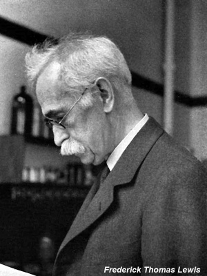
Paper - The gross anatomy of a 12 mm pig
| Embryology - 28 Apr 2024 |
|---|
| Google Translate - select your language from the list shown below (this will open a new external page) |
|
العربية | català | 中文 | 中國傳統的 | français | Deutsche | עִברִית | हिंदी | bahasa Indonesia | italiano | 日本語 | 한국어 | မြန်မာ | Pilipino | Polskie | português | ਪੰਜਾਬੀ ਦੇ | Română | русский | Español | Swahili | Svensk | ไทย | Türkçe | اردو | ייִדיש | Tiếng Việt These external translations are automated and may not be accurate. (More? About Translations) |
Lewis FT. The gross anatomy of a 12 mm pig. (1903) Amer. J Anat. 2: 221-225.
| Online Editor |
|---|
| This historic 1903 paper by Lewis described development of the early pig.
|
| Historic Disclaimer - information about historic embryology pages |
|---|
| Pages where the terms "Historic" (textbooks, papers, people, recommendations) appear on this site, and sections within pages where this disclaimer appears, indicate that the content and scientific understanding are specific to the time of publication. This means that while some scientific descriptions are still accurate, the terminology and interpretation of the developmental mechanisms reflect the understanding at the time of original publication and those of the preceding periods, these terms, interpretations and recommendations may not reflect our current scientific understanding. (More? Embryology History | Historic Embryology Papers) |
The Gross Anatomy of a 12 mm Pig
By
Frederic T. Lewis, A. M., M. D.
From the Embryological Laboratory, Harvard Medical School. With 4 Plates.
Introduction
It has been my privilege to study, with Prof. Charles S. Minot, the transverse sections of a 12-mni. pig embryo forming Series 5 of the Harvard Collection. Numerous sections of this embryo are to appear in Prof. Minot's new "Laboratory Text-book of Embryology." To show the relation of these to one another I have made four reconstructions which are here separately presented as the basis of this paper. It is thought that this particular pig is distinguished from all other embryos of corresponding size by the detail and completeness with which the entire animal has been portrayed. Similar figures of the human embryo are found in PI. XX, Vol. 5 of the Journal of Morphology (Mall, 91), and in Taf. II of His's contribution " Zur Geschichte del Gehirns," 88.
The reconstructions were made generally by the method of His. The contour of the brain and pharynx was taken from wax models prepared by Dr. J. L. Bremer; elsewhere the shading has been inferred from studying the cross sections. Since the umbilical cord was cut away in the embryo of series 5, that region was supplied from another transverse series of a 12-mm. pig, No. 518 of the Har\'ard Collection.
The general description of the illustrations is left to the lettered figures. Only the more interesting features are presently to be described ; first, those concerning the brain, nerves, and sense organs ; then those involving the pharynx, digestive tract, and viscera ; finally, those relating to the vascular system.
Brain, Nerves, and Sense Organs
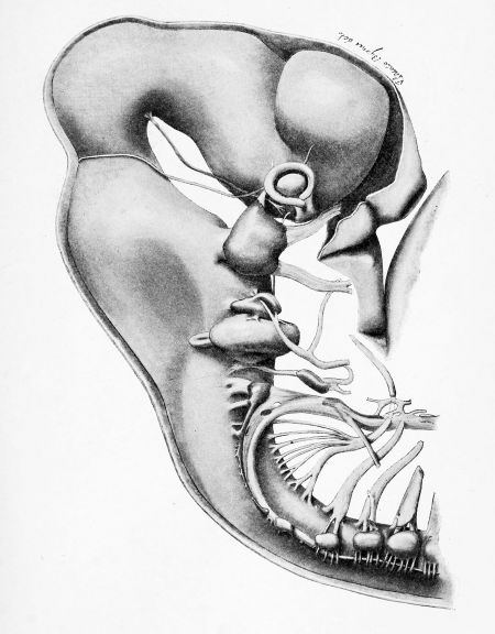
The exterior of the brain and nerves appears in Plate I; the internal aspect of the brain in Plate III; and the internal view of certain nerves in Plate IV. Unfortunately the depression between mid-brain and diencephalon has been exaggerated in some of the figures. In the median plane ,the superficial surface is not indented at this point. The rounded outer wall of the diencephalon shows no trace of the epiphysis which begins to form in a pig of 20 mm. and becomes well defined in one of 24 mm. The hind brain possesses three well marked nenromeres followed posteriorly by a fourth shallow one. In a 1) mm. pig there are five which are distinct.
Of the nerves shown in Plate I, the fifth is noteworthy as possessing no ciliar}' ganglion. Its ophthalmic division is not connected with either the third or fourth nerves, though it passes very near the latter. It finally divides into a shorter frontal and a longer nasal branch (Dixon). Ciliary ganglia have been drawn as if belonging with the fifth nerve by both His and Mall, but as shown by Dixon, 96, and admitted by Prof. His, this was an error. No ciliary ganglion could be found in this pig. The motor tract of the fifth nerve passing into the inferior maxillary division is drawn in Plate IV.
The seventh nerve, Plate I, appears as a bundle of fibres free from cells passing out from the lower part of the lateral wall of the medulla. Its course within the brain proceeds from a point near the median line directly outward, transverse to the cerel)ral axis. Outside of the medulla a large ganglionic mass is placed over the motor bundle in equitant fashion. That part toward the fifth nerve is the geniculate ganglion from the inferior end of which passes a clump of fibres to fuse with the motor bundle already described, and form the main trunk of the facial nerve. The geniculate ganglion is in close contact with the acoustic ganglion and in a few sections is inseparable from it. From the geniculate ganglion there arises a slender fasciculus, the pars intermedia of Wrisberg. It proceeds backward under the entering root of the eighth nerve, to the wall of the medulla, Plate IV. After penetrating into the brain, the fibres lie just ventral to the oval bundle, and may be traced a short distance caudad, running parallel with the cerebral axis. Duval has followed them to the upper end of the glosso-pharyngeal nucleus (Quain). The slender pars intermedia, the large geniculate ganglion and the motor root are parts of a single nerve, secondarily united with the acoustic ganglion. Eesearches establishing this opinion are cited by Thane, 95, Van Gehuchten, 97, and WiedersJieim, 02. The further course of the nerve is shown in Plate 1. It divides into prae-trematic and post-trematic branches, but the division is under the spiracle or auditory cleft and not over it as in fishes. The anterior branch of the seventh nerve passes into the mandibular arch as the chorda tympani; the remainder lies behind the auditory groove in the hyoid arch.
The eighth nerve shows its upper vestibular and lower cochlear branches, but its ganglion is not yet subdivided.
The trunk of the ninth nerve is cellular for some distance before it enters the medulla, but possesses no distinct ganglion other than the ganglion petrosmn. Just beyond the ganglion a very slender branch, not drawn in the figure, passes upw-ard and forward over the second cleft. This is Jacobson^s nerve. The main stem passes down behind the cleft Avhere it is seen dividing into the lingual branch which extends forward into the hyoid arch, and a smaller pharyngeal branch which remains in the third arch. Notwithstanding the changed relations to the second cleft (such as has occurred in the seventh nerve) the lingual branch is presumably comparable with the prae-trematic and the pharyngeal with the post-trematic of fishes. In this embryo there was no connection betAveen the ninth and tenth nerves.
The tenth nerve arises from the large jugular ganglion, extending from which is a beaded commissure ending in a small knob. In the track of the commissure, but separated from it, and lying beyond it, is an irregular ganglionic mass. After another interval there appears a small fragment, and then follow^s the first cervical ganglion notably smaller and more dorsally placed than those which succeed it. The irregular ganglionic mass is not connected with the hypoglossal nerve in series 5, but in series 518 a slender bundle unites the two structures. This, then, is Froriep's ganglion. Its relation with the commissure is far more striking than its resemblance with a spinal ganglion. I have found it connected with the commissure in pigs of 17 mm., as did Froriep in a sheep of 12.5 mm. He considered the connection unimportant and described the " vagus ganglion " as passing beneath the hypoglossal ganglion. In his figures (82, PL 16) the latter is drawn of the same form and texture as the spinal ganglia, and very different from the diffuse jDrolongation of the vagus ganglion. In a dissected pig of 17 mm. I could find no such difference: the hypoglossal ganglion api>eared as a detached part of the ganglionic, chain running forward to the vagus. This commissure in 17 mm. embryos could not be subdivided into definite ganglia; it w^as characterized by irregular SAvel lings and spurs. In the adult it remains as one or two hypoglossal ganglia. Froriep and Beck, 95, p. 689, in all of the six hogs examined, found a single hypoglossal ganglion and in one case two. In man they are quite constantly absent and the degeneration extends to the first cervical ganglion which may even be macroscopically lacking.
Below the ganglion nodosum the vagus nerve gives rise to several branches passing betw^een the third and fourth branchial clefts. One branch passes under the third cleft into the arch in front of it. There is a branch behind the fourth cleft and the system ends with the large recurrent larjugeal nerve. The laryngeal plexus appears to be derived from the nerves to the degenerated gill clefts.
The spinal accessory nerve begins at the level of the sixth cervical ganglion. Its fibres are very closely associated with the hypoglossal ganglia; in series 518 it rests against, and in places is nearly surrounded l)y the first cervical ganglion. The passage of fibres between the latter and the spinal accessory nerve has been found in man by Kassander^, and con^rmed by Froriep and Beck, 95 (p. 694).
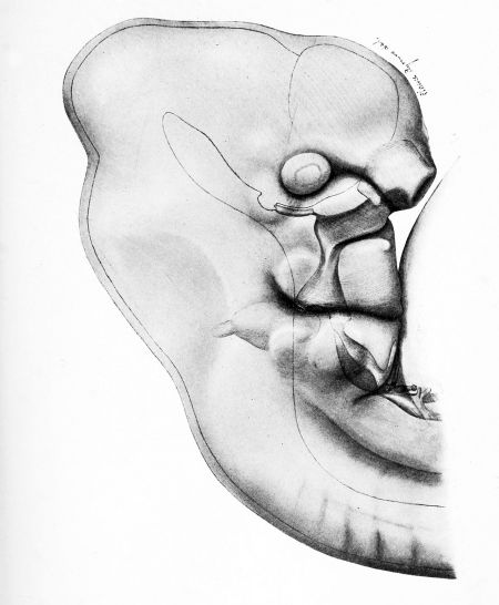
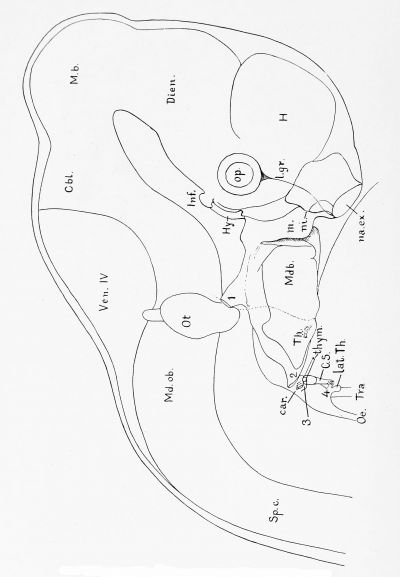
Of the sense organs, only the nasal cavity need be noted. Plate 2 is drawn to show the relations of the pharynx and some other internal structures to the surface markings. The nasal cavity appears with the external naris opening broadly on the surface. Toward the mouth the two edges of this cavity are brought together by the growth of the median nasal process on the inside, and of the lateral nasal and maxillary processes (separated by the lachrymal groove) on the outside. Caught between the internal nasal and maxillary processes, the walls of the nasal opening are compressed to form a raphe of semicircular outline as shown in the figure. This raphe extends to the roof of the oral cavity. At its internal end it is a mere plate, indicating the position of the internal naris. In the 14-nim. pig the raphe has disintegrated and mesenchyma extends from side to side. The membrane is at is internal end, after becoming broad and thin, ruptures. Hochstetter, 92, states that " the primary choana? of mammals arise from a breaking through of the hind end of the nasal pit by a tearing apart of the membrana bucconasalis, and there exists in mammals no primary connection between the nasal and buccal cavities." Peter, 02, pp. 5455, adopts this interpretation. The new opening, however, appears to involve only a part of the raphe made by the fused lips of the primitive nasal opening. The fusion is permanent except in the region of the from the entodermal tract.
Pharynx, Digestive Tract, and Viscera
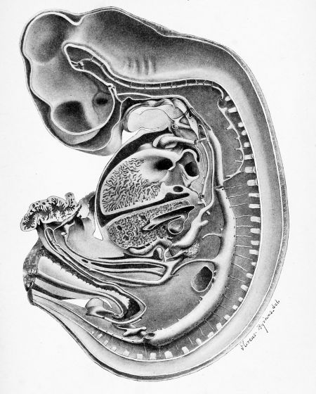
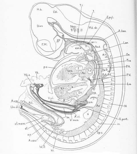
The structures connected with the roof of the mouth are shown in Plate II, and in median section also in Plate III. Beginning anteriorly there first appears the hypophysis, having a slender outlet and a broad, thin, spade-like body, flattened parallel with the brain wall. It tends to fork at the infundibular gland and possesses an inconstant knob near the junction of its body and duct. Behind the duct there is still to be seen Seessel's pocket; between it and the hypophysis the oral membrane formerly separated the stomadaeum from the entodermal tract.
The floor of the oral cavity is shown in relief in Plate III. Most anteriorly are the large mandibular arches. From the broad dorsum of each arises an elongated eminence, rounded in cross section. In the median line between them, there is a groove in the caudad part of which is found a low elevation, the tubereuhini inipar of His. Kallius, 01, p. 43, has named this pair of mandibular elevations the lingual folds, and states that they form almost the entire body and tip of the tongue. The tuberculum impar is the source of the posterior part of the lingual l)ody and of the septum. \ shallow transverse groove still marks the fusion of tuberculuui impar and the root of the tongue. The thyroid duct which opened into this groove has become obliterated, and the circumvallate papillae which are to develop along its course have not yet appeared. That the root of the tongue is formed by a fusion of the second and third arches, as stated by His, 85, p. G5, could hardly be determined at this stage.
Behind the tongue there is a short, low, l)ilol)ate protuberance, the epiglottis. Beyond this the ventral will of the pharynx presents a round, dorsal ly directed elevation, which soon becomes indented in the median line. Thick conical masses, the arytenoid folds, appear on either side of this groove. The notch between them becomes a long, slender slit. The trachea, after separating from the oesophagus, continues in this slit-like form, its lateral walls being in close contact except at their dorsal end, where a minute passage exists. Further caudad the ventral prolongation disappears, and the trachea is then a simple tube, Plate 111. The subsequent development of the larynx has not been adequately described.
The oral cleft, forniing one of a pair of lateral wings at the beginning of the digestive tract, is represented in Plate II. The auditory cleft rises dorsal to the pharyngeal wall, and passes to the auditory groove. It does not open to the exterior. Its internal orifice is drawn in Plate IV, in which it is seen to end behind a conical mass pendant from the side of the pharyngeal roof. The anterior part of the Eustachian opening meets the beginning of the oral wing.
The second pharyngeal pouch runs posteriorly outward, parallel with the course of the pharynx. It opens freely into a well marked external groove which subsequently deepens and separates the lower jaw from the neck. There is no indication of a " Verschlussplatte " in series 5 ; in series 518, an oblique plate is present on both sides, but appears to be broken through. In a 10-mm. pig, the clefts opened to the exterior but remnants of the plates existed. As yet no indication of the tonsils appears in connection with the second cleft.
The third pouch is a slender tube passing outward, at right angles with the pharynx to its small " Verschlussplatte." Just within the plate it gives rise to a slender tube passing ventrally, parallel with the external groove of the second cleft. This diverticulum may represent an elongated contact with the ectoderm, since its course is parallel with the corresponding part of the second cleft. De Meuron, 86, p. 10-i, states that in ho class of mammals other than vertebrates can anything be found comparable with this ventral coecum.
The closing plate of the third cleft lies very near a deep ectodermal pocket which opens externally by a clear-cut nearly round hole, and extends internally along the course of the tenth nerve, ending in an epithelial proliferation. A very slender tube from the pharynx runs toward this mass. The pocket, gland and tube mark the course of the fourth cleft.
From the base of the entodermal part of the fourth cleft there is another pocket tending to pass around in front of the trachea. De Meuron considered this to be a rudimentary cleft. It may, however, be a ventral branch of the fourth cleft comparable with that of the third. Both of these ventral processes occupy ]3arallel positions in the embryo.
In a 12-mm. pig, the cervical sinus is mainly the ectodermal part of the fourth cleft as above described. Its rounded opening has not yet been concealed by the opercular extension of the hyoid arch. The closing plate of the third cleft is becoming involved in the orifice of the sinus anteriorly. Posteriorly the outlet of the sinus is in contact with the ganglion nodosum forming Froriep's epibranchial organ, 85. The epithelial jH'oliferation shown in Plate II is below the level of the ganglion.
The thyroid gland is represented in the 12-mm. pig by its somewhat branched median anlage in the second arch, and by the ventral arms of the fourth cleft. The latter, known also as the post-branchial bodies or lateral thyroids, encircle the trachea, become detached from the pharynx, and, in the higher mammals only, connect with the median thyroid.
The thymus is derived mainly from the ventral arm of the third pouch which also encircles the trachea, approaching its mate from the opposite side. In the 12-mm. pig there is an epithelial mass connected with the entoderm of the third arch and with the adjacent ectoderm. In the 14-mm. pig, this tissue has apparently fused with that at the tip of the cervical sinus, and the resulting mass is also in contact with the ganglion nodosum. Froriep, 85, p. 32, found that his study of the epibranchial organ was complicated inasmuch as the cell mass, connected on one side with the ganglion of the tenth nerve, was united on the other with the anlage of the thymus. Such a condition appears in a 17-ram. pig. De Meuron, 86, p. 76, found that in sheep the fourth arch produced a structure similar to the superior part of the thymus but which remained behind the thyroid gland. As already stated, in the 1-i-mm. pig this mass has apparently united with the superior part of the thymus. In an embryo of 2i mm. the thymus consists of the approximated and proliferated ends of the ventral arms of the third clefts. From each of these a slender cord passes upward and backward to join the considerable cell mass near the vagus. This connection is broken, and the small upper part has been variously called the carotid gland, the parathyroid body or, more recently, the epithelial body (Maurer, 02).
The right lung is figured in Plate III. Its elongated condition as compared with the human lung is noteworthy. Above the bifurcation of the trachea there is a budding bronchus which is to supply the upper lobe of the right lung. No corresponding branch is found on the opposite side. This asymmetry of the lungs, described by Aeby, has been found well developed in the lowest of existing mammalia (Wiedersheim, 02, p. 434). With this, its early embryonic appearance is in full accord. Keibel, 97, found the tracheal bronchus in a 9.6-mm. embryo; in the Harvard Collection it is well developed at 9.0 mm.
The stomach is already a pig's stomach, possessing in its cardiac portion a well marked diverticulum.
The liver comprises four large lobes which are visible before the embryo is sectioned. Along its lower surface there extends a pouch, exceeding in diameter the intestine from which it arises and into which it empties. Its cylindrical epithelium is quite unlike that of the hepatic cylinders, but resembles somewhat the intestinal mucosa. This large diverticulum ends blindly, its terminal portion being separated from the liver by mesenchyma. Sometimes a knob-like bud is found on its surface. JvTearer the intestine these buds are more numerous. Some of them terminate in hepatic cylinders ; others end blindly, or are found as detached cysts in the liver. Later all but one of these hepatic ducts are obliterated, and the diverticulum into which it empties has become in part the cystic duct and in part the gall-bladder. Brachet, 96, p. 666, describes similarly the development of the rabbit's biliary ducts. There is first a single intestinal diverticulum, from the proximal two-thirds of which the hepatic cylinders proliferate, and of which the quiescent distal part is retained as the gall-bladder. From its em])ryological history, it is to be expected that the hepatic ducts possess glands " resembling those of the gastric cardia " and that in the distended gall-bladder such glands should be absent.
The pancreas consists of two closely applied divisions. The ventral milage empties into the bile duct close to the intestine. The dcn-sal and larger portion, separated from the ventral by the portal vein, opens into the duodenum below the bile duct. The adult pancreas is formed of a band of gland-tissue extending from the biliary duct to the duct of Santorini, which opens into the intestine 15 cms. lower down." From this band extend two tails which seem to correspond with the two anlages. The ventral duct, that of Wirsung, is represented by impervious fibrous tissue. The duet of Santorini, from the dorsal anlage, is the only one retained in the adult pig.
The intestine extends into the umbilical cord as a simple loop. After receiving the yolk stalk which is now very slender and scarcely pervious, it returns to the body. A dilatation marks the position of the coecum, which in the adult is a voluminous pouch without an appendix.
The rectum, just before entering the cloaca, is nearly occluded by an epithelial proliferation which separates the intestinal and urinary tracts. In a 20-mm. pig the anus is distinct from the urogenital sinus, and the proliferation has wholly disappeared. I have found a similar plug in rabbits, and Keibel, 96, has figured one in an ll.5 mm. human embryo. He describes it as an "accidental and insignificant adhesion." Gasser discovered a corresponding structure in the chick which has been fully described and its function explained by Minot, 00, 2.
The urogenital system consists of very large Wolffian bodies outlined in Plate lY, and of the genital ridges. The kidneys are represented by their pelves, Plate III. From the latter, the slender ureters pass to the Wolffian ducts, which in turn unite witli the allantois and enter the cloaca.
The cloaca is closed by its membrane, or rather by the approximation of its borders which form a raphe. This is to open at its ends, giving rise to the anal and urogenital apertures, and to remain fused between them, forming the perineal raphe.
Heart, Arteries, and Veins
The heart, because of its trabecular structure, must be drawn diagrammatically. In Plate III it is shown cut through on the left side of the median septa. The constriction between the auricles forms a partial septum. Below this and toward the left side of the embryo is the large foramen ovale, bounded partly by the auricular wall and partly by the thin wavy septum superius. This septum is attached ventrally to a thick mesenchymal mass, from which proceeds the shelf-like anlage of the atrio-ventricular wall and mitral valve. The left auricle passes caudad into a funnel-like prolongation, the pulmonary vein. As in -man, this is at first a single tube, but later is taken up into the auricle so that its several branches enter by separate orifices.
- In man, His (85, pp. 19 and 34) has tisured the dorsal anlage in its later stages as emptying below the bile duct, as in the pig. Hamburger, Schirmer and others tind the duct of Santorini opening above the bile duct.
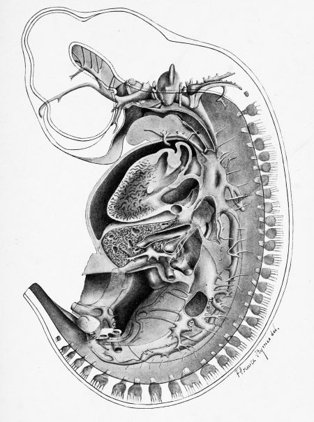
The left ventricle is cut off from the right by a trabecular partition, capped dorsally by a mass of dense mesenchyma which bounds the illnamed interventricular foramen. This foramen opens into the space a. 1). c, shown in a section of the heart on the right of the median septa, Plate lY. From here the blood may proceed eitlier through the aorta or the pulmonary artery. The aorta and pulmonary artery are separated by a pair of folds united above, but distinct beloAv. The fold on the left side, which has been cut away, passed over into the interventricular septum near b. The other extends along the right cardiac wall, ending in the tricuspid anlage. These folds fuse so as to connect a with b, and b with c. The only outlet for the persistent interventricular foramen is then into the aorta (Born, 89, p. 339). The cut tissue near h is not involved in the division of the bulbus arteriosus just described. It represents a clump of trabecula? passing from the right wall and sectioned just before uniting with the median septum. It is connected with the tricuspid trabecula?. Instructive cross-sections at this level have been drawn by Hochstetter, 02, p. 55.
The right auricle still has a single opening for the ducts of Cuvier and the inferior vena cava. They unite in the sinus venosus which empties between its two valves. The valves are united above, forming the septum spurium. In man the septum spurium and a part of the left valve are used in closing the foramen ovale; parts of the right valve persist as the Eustachian valve and valve of Thebesius.
Arteries
The arterial system is represented in Plate III. The pulmonary artery leaves the heart by a single trunk, which divides into two arches of equal calibre, passing to the right and left aort« respectively. From the right arch a large stem, and from the left a slender one, unite and proceed to the lungs. Only the left arch and its stem contribute to the adult pulmonary artery as described by Bremer, 02.
The aorta also begins as a single trunk wdiich bifurcates, forming right and left divisions. Beyond the bifurcation the ventral section of the aorta continues to the tong-ue and jaw as the external carotid artery. External and internal carotid arteries are united by the carotid arch. A very short section of ventral aorta represents the common carotid artery.
Proceeding caudad, the internal carotid artery passes into a slender degenerating portion of the dorsal aorta extending to the aortic arch, beyond which it becomes a large trunk which unites with a similar vessel from the oiDposite side to form the median dorsal aorta.
From the median aorta there arise pairs of intersegmental arteries. These formerly continued in single column along the right and left aortte, but after the development of the vertebral anastomosis they lose their aortic origin. In the 12-mm. pig the seventh intersegmental artery is the first to arise from the right aorta. From it the vertebral and subclavian arteries originate.
The subsequent development of these vessels has been figured by Eathke, 43. The minute common carotid arteries become very long stems and fuse with one another below. Xear their junction arises the right subclavian, of which the right aorta down to the 7tli intersegmental artery forms a small section. Common carotids and right subclavian come from an innominate artery, not yet formed in the 12-mm. pig. The left subclavian leaves the aorta separately. Caudad from the 7th intersegmental artery, the right aorta is obliterated.
The arteries of the brain have nearly their adult arrangement. The vertebrals unite in a long basilar artery w^th many lateral branches. In front of the pons it forks, forming the posterior communicating arteries, which pass into the internal carotids. A branch extending between optic stalks and brain forms the anterior cerebral artery which by anastomosing with the opposite side will complete the circle of Willis. In front of the mid-brain are the posterior cerebral arteries.
The branches of the dorsal aorta are the regular pairs of intersegmental arteries, the scattered mesonephric arteries and, in the median line, the coeliac axis and the omphalo-mesenteric artery. The last named formerly encircled the intestine ( Hochstetter, 02, p. 114), but the left half of the loop has disappeared, and it now passes to the yolk sac on the right of the intestine. The large umbilical Ijranches and the caudal extension of the aorta complete the arterial system.
Veins
The veins are shown chiefly in Plate IV. The anterior cardinal system arises as a plexus between the developing hemispheres. The small vessels unite in a median stem, the anlage of the superior longitudinal sinus. This soon divides into two branches which pass around to the under side of the brain, thus forecasting the lateral sinuses. It receives the ophthalmic vein, later subdivided into ophthalmic vein and cavernous sinus. ]\Iany branches from the mid-brain region unite with those just described and form the internal jugular vein. The 12-mm. pig agrees very closely with the 6-mm. guinea-pig studied by Salzer, 95.
The internal jugular vein passes between the Gasserian ganglion and the l)rain ; thence outside of the otocyst, and nerves seven to eleven inchisive, finally becoming internal to the twelfth nerve near the ganglion nodosum. Originally internal to all these nerves, it later becomes external to them all (Salzer).
A vein coining from the side of the tongue passes outside of the tenth nerve and enters the internal jugular at the level of the lateral thyroid pocket. The corresponding vein from the left side begins in the median line, along which it continues a short distance before turning to one side. In a 14-mm. pig the right vein is the median one. Anastomosis between the two is probable. These vessels, which to ray knowledge have never been described, are found in 6-mm. pigs, running along the dorsal pericardial wall and proceeding from as far forward as the second arch. In a pig of 30 mm. the anterior cardinal veins have approached so near that the intervening space is less than the diameter of one of them. Where they are nearest these transverse veins are found, passing ventral to the arteries, dorsal to the cords of the thymus, and just caudad from the thyroid, with which they are closely connected. Here the very short left innominate vein is formed, uniting the anterior cardinals. That part of the right cardinal between the anastomosis and the heart subsequently disappears.
Near the duct of Cuvier the anterior cardinal vein is split for some distance, and the subclavian vein arises from its outer part. That section of the external jugular which is near the heart appears to be cut off from the internal jugular by the growth of slender mesenchymal partitions. After the disappearance of the posterior cardinal vein, the right duct of Cuvier becomes a continuation of the internal jugular vein, and with the migration of the subclavian vein, adult conditions ensue. Shortly before birth, these veins are arranged like a four-tined fork, the handle being the vena cava superior (duct of Cuvier and anterior cardinal), the inner tines being the internal jugulars (anterior cardinals), and the outer tines representing the external jugulars from which the subclavians pass off laterally.
The posterior cardinal veins arise near the tail and enter the Wolffian bodies where they become divided into sinusoids (Minot, oo, 1). They form distinct trunks as far cephalad as the cross anastomosis between the subcardinal veins. This section of the right posterior cardinal becomes a part of the inferior cava, the corresponding portion on the left forms a part of left spermatic vein. A prominent anterior trunk unites with two others and proceeds to the duct of Cuvier. This section of the posterior cardinal vein loses its connection with the heart on the right side, but retains it on the left, forming the hemiazygos vein. The fusion of azygos and hemiazygos is described by Parker and J'ozier, 98.
The subeardinal veins, Lewis, 02, are formed by an anastomosis of Wolffian sinusoids. The eephalad portion of tlie right subeardinal conveys the blood from posterior cardinal to the liver, and is a part of the inferior vena cava. That on the left disappears.
The umbilical veins, which in the cord of series 518 are fusing with one another, separate in the embryo and extend through the body wall to the liver. Here the left umbilical vein is broken into sinusoids except for a quite open passage to the portal vein. The larger right umbilical vein passes through the liver as the ductus venosus Arantii which joins with the portal vein and inferior vena cava to form the vena hepatica communis.
It remains to describe the portal vein, the hepatic relations of which appear in Plate lY. It is a continuation of the omphalo-mesenteric vein as shown in Plate III. That vein, beginning with branches from the yolk sac, crosses the abdominal cavity in a detached bit of mesentery and passes along the left side of the duodenum where it receives the superior mesenteric vein. The mesenteric vein begins in the intestinal loop, not in the yolk sac. Similar conditions have been found in the cat by Dexter, 02, from whose work it appears that the mesenteric vein is a new branch, and not one of the oniplialo-mr'senteric vessels, as has generally been supposed.
Prof. Minot has shown in his laboratory text-book that the thorough study of a few embryos is for students the shortest Avay to a comprehensive knowledge of embryology. As a means for correlating special embryological investigations, the complete description of single embryos is a promising but almost untried experiment.
Note. — It should be understood that thoug'h the results here presented have been drawn upon in part in the making of a text-book, the present study is complete in itself and independent of the text-book. This paper accepted for publication in Xo. 1, Vol. 2. was unavoidably delayed till now by circumstances beyond the control of the aiithor. — Editor.
Publications Cited
Bkachet, a., g6. — Le developpement du pancreas et du foie. Journ. de I'Anat. et de la Physiol., Vol. 32, pp. 620-696.
Bremer, J. L., 02. — On the origin of the pulmonary arteries in mammals. Amer. Journ. of Anat., Vol. 1, pp. 137-144.
BoRX, G., 89. — Beitrage zur Entwicklungsgeschichte des Saugethierherzens. Arch. f. mikros. Anat., Bd. 33, pp. 283-378.
Dexter, F., 02. — On the vitelline vein of the cat. Amer. Journ. of Anat., Vol. 1, pp. 261-267.
Dixon, A. F., g6. — On the development of the branches of the fifth cranial nerve in man. Trans, of the Boy, Dublin Soc, Vol. 6, Pt. 2, pp. 19-76.
Frobiep, a., 82. — Ueber ein Ganglion des Hypoglossus. Arch. f. Anat. u. I'hys.; Anat. Abth., pp. 279-302.
Ss- — Ueber Anlagen von Sinnesorg'anen am Facialis, Glossopharyngeus und Vagus. Arch. f. Anat. u. Phys.; Anat. Abth., pp. 1-55.
Fkokiep, a., und Beck, W., 95. — Ueber das Vorkommen dorsaler Hypoglos suswurzeln mit Ganglion in der Eeihe der Siiugetiere. Anat. Anz., Bd. 10, pp. 688-696. Van Gehuchten, A., 97. — S3^steme nerveux de I'homme. 2nd ed., pp. 1-941.'
His, W., 85. — Anatomie menschlicher Embryonen, iii, pp. 1-260.
88. — Zur Geschichte des Gehirns. Abbl. d. Math.-phys. Classe der K.
Sachs. Ges. der Wiss., Bd. 14, pp. 343-392.
HocHSTETTER, F., 92.— Ucbcr die Bildung der primitiven Choanen beim Menschen. Verh. der Anat. Ges., 6, pp. 181-183.
02. — Die Entwickelung des Blutgefasssystems. Handbuch d. verg. u. exp. Entw. der Wirbelthiere, Bd. 3, pp. 21-166. Kallius, E., 01. — Beitrage zur Entwickelung der Zunge. Verh. der Anat. Ges., 15, pp. 41-42.
Keibel, F., 96. — Zur Entwickelungsg'eschichte des menschlichen Urogeni talapparates. Arch. f. Anat. u. Phys.; Anat. Abth., pp. 55-156.
97.— Nomientafel 1. Zur Entwicklungsgeschichte des Schweines, pp. 1-113.
Lewis, F. T., 02. — The development of the vena cava inferior. Amer. Journ. of Anat., Vol. 1., pp. 229-244.
Mall, F., 91. — A human embryo twenty-six days old. Journ. of Morph., Vol. 5, pp. 459-478.
Maurer, F., 02. — Die Entwickelung des Darinsystenis. Handbuch d. vergl. u. exp. Entw. der Wirbelthiere, Bd. 2, pp. 109-252. De Meubon, p., 86. — Le developpement du thymus et de la glande thyroide.
MiNOT, C. S., 00, 1. — On a hitherto unrecognized form of blood circulation. Proc. of the Bost. Soc. of Nat. Hist., Vol. 29, pp. 185-215. 00, 2. — On the solid stage of the large intestine in the chick. Journ. of the Bost. Soc. of Nat. Hist., Vol. 4, pp. 153-164.
Parker, G. H., and Tozier, C. H., 98. — The thoracic derivatives of the postcardinal veins in swine. Bull, of the Mus. of Comp. Zool., Vol. 31, pp. 133-144.
Peter, K., 02. — Die Entwickelung des Geruchsorgans, u. s. w. Handbuch d. vergl. u. exp. Entw. der Wirbelthiere, Bd. 2, pp. 1-82. Katuke, H., 43.^ — Ueber die Entwickelung der Arterien welche bei den Siiugethieren von dem Bogen der Aorta ausgehen. Miiller's Archiv, pp. 276-302.
Salzeb, H., 95, — Ueber die Entwicklung der Kopfvenen des Meerschwein chens. Morph. Jahrb., Bd. 23, pp. 232-255.
TiiANE, G. D., 96.— The Nerves. Quain's Anatomy, Vol. 3, Pt. 2, pp. 221-403. Wiedersheim, R., 02. — Vergleicheude Anatomic der Wirbelthiere, 5th ed , pp. 1-686.
Explanation of Plates
Plate I
|
|
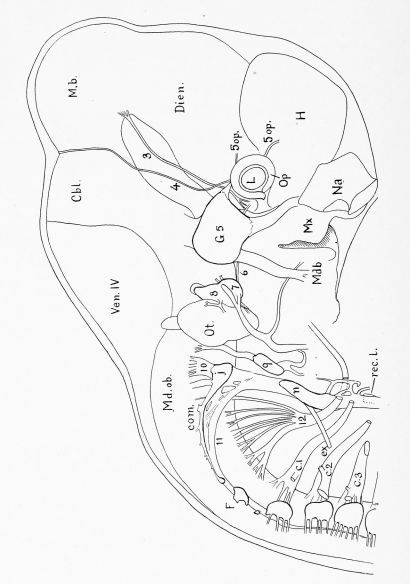
Pig Embryo of 12.0 mm. Reconstruction from transverse sections, Series 5.
To show especially the cephalic nerves, c. 1, c. 2, c. 3, Cervical nerves. Cbl, Cerebellum, com. Ganglionic commissure. Dien, Diencephalon. e.i'. External branch of the spinal accessory nerve. F, Froriep's ganglion. Gf. 5, Gasserian ganglion. H, Cerebral hemisphere. ;, Jugular ganglion. L, Lens. M. b, Mid-brain. Mdb., Mandibular process. Md. ob, Medulla oblongata. Mic, Maxillary process, n, Ganglion nodosum. Na, Nasal pit. Op, Optic cup. Ot, Otocyst. Rec. I, Recurrent laryngeal nerve. Yen. IV, Roof of fourth ventricle. 3, Oculomotor nerve. 4, Trochlear nerve. Sop, Branches of the ophthalmic division of the trigeminal nerve. 6, Abducens nerve. 7, Geniciilate ganglion of the facial nerve. 8, Vestibular ganglion. 9, Petrosal ganglion. 10, Vagus nerve. 11, Spinal accessory nerve. 12, Hypoglossal nerve. x 20 diams.
Plate II
|
|
Pig Embryo of 12.0 mm. Reconstruction from transverse sections, Series 5.
The embryo has been drawn as if transparent to show the form of its pharj'^nx, and the relations of the pharyngeal gill pouches to the grooves on the outer surface of the embryo, car. Carotid gland. Cbl, Cerebellum. CS, Cervical sinus. Dieii, Diencephalon. H, Cerebral hemisphere. Hi/, Hypophysis, liif. Infundibular gland. Lat. Th, Lateral thj^roid. 1. gr. Lachrymal groove, m. Maxillary process. M. b. Mid-brain. Mdb, Mandibular process. Md. ob. Medulla oblongata, na. ex. External naris. ni, Internal naris, closed by an epithelial plate. Oe, Oesophagvis. Op, Eye. Ot, Otocyst. Sp. c. Spinal cord. Th, Median thyroid gland, thym. Thymus. Tra, Trachea. 1, 2, 3, Ji, Entodermal pouches of the corresponding gill clefts. x 20 diams.
Plate III
|
|
Pig Embryo of 12.0 mm. Reconstruction from transverse sections, Series 5 and 518.
For the most part the organs represented are in or near the mediae plane. The drawing illustrates especially the alimentary tract, the arterial system, and the heart. A. bas. Basilar artery. A. can. Caudal artery. AU, Allantois. Ao, Median dorsal aorta. An. d, Ao. D, Right dorsal aorta. A. s. Subclavian artery. Am, Left auricle. A. um, Umbilical artery. A. ti,. Vitelline artery (Omphalo-mesenteric). c, Coeliac axis. car. e. External carotid artery, car. i. Internal carotid artery. Cbl, Cerebellum, cl. Cloaca, cl. mem, Cloacal membrane, d. Left duct of Cuvier. Dion, Diencephalon. D. V, Ductus venosus Arantii. ep, Epithelial plug in the rectum. Epgl, Epiglottis. f, interventricular foramen opening into the space a, b, c, of PL 4. Fni, Foramen of Monro, f, o. Foramen ovale, g, Gall-bladder. In, Entodermal wall of the intestine, is, An intersegmental artery. Ki, Renal pelvis. Lar, Larynx, Li, Liver. Lu, Lung. M. b. Mid-brain. Md. ob. Medulla oblangata. N, A spinal nerve. Neu, Neuromeres. Oe, Oesophagus, op. Optic stalk. P. A, Pulmonary artery. Pan, Dorsal anlage of the pancreas, behind the duct of which is the ventral anlarge. P, c. Peri-cardial cavity, P. V, Pulmonary vein. Sp. c, Spinal cord. St, Stomach. /, Posterior portion of the tongue. T. /, Tuberciilum impar. Ti'u, Trachea. Um. d, Eight iimbilical vein. Vcn, Left ventricle. T. mes, Superior mesenteric vein. V. port. Portal vein. Y. in. Vitelline vein (Omphalo-mesenteric. W. D, Wolffian duct, x. Anastomosis between the right and left cardinal systems. Yk, Yolk sac. Yfc. s, Yolk stalk. x 13.5 diams.
Plate IV
|
|
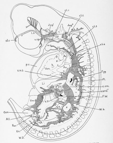
Pig Embryo of 12.0 mm. Reconstruction from transverse sections, Series 5.
The section is on the right of the median plane, and shows chiefly the heart and venous system. 1, Umbilical artery, a. Aortic septum. All, Allantois. Ao, Aorta. Au, Eight auricle. 6, Bundle of trabeculae. c, wall between bulbs arteriosus and auricle, card', card". Superior and inferior sections of the posterior cardinal vein. cl. 1, cl. 2, cl. 3, cl. ^, Entodermal poiiches and their pharyngeal openings of the corresponding gill clefts, c. am. Dotted outline of the omental or lesser jjeritoneal cavity, d, Left duct of Cuvier. D. C, Eight duct of Cuvier. D. V, Dvictus venosus Arantii. F. ^V, Foramen of Winslow. F. pp, Pleuro-peritoneal foramen, gen. Genital txvbercle. G. R, Genital Eidge. Jug', Jug", Jugular or anterior cardinal vein. Li, Liver. /. s, anlage of the lateral sinus, mx, Transverse vein. P. Pulmonary artery, p. c. Pericardial cavity. PI, Dotted outline of pleural cavity, liec, Eectum. Sc, Subcardinal vein. Scl, Subclavian vein, sis, Anlage of the superior longitudinal sinus. Ur, Ureter. Um. d. Eight umbilical vein. r. Valves of the sinus venosus. Yen, Eight ventricle. Y. H. C, Vena hepatica communis, v. op. Ophthalmic vein. Y. P, Portal vein. W. ft, Wolffian body. W. D, WolflTian duct, x. Anastomosis between the right and left cardinal systems. x 13.5 diams.
Cite this page: Hill, M.A. (2024, April 28) Embryology Paper - The gross anatomy of a 12 mm pig. Retrieved from https://embryology.med.unsw.edu.au/embryology/index.php/Paper_-_The_gross_anatomy_of_a_12_mm_pig
- © Dr Mark Hill 2024, UNSW Embryology ISBN: 978 0 7334 2609 4 - UNSW CRICOS Provider Code No. 00098G


