Paper - The Earliest Blood-Vessels in Man
| Embryology - 27 Apr 2024 |
|---|
| Google Translate - select your language from the list shown below (this will open a new external page) |
|
العربية | català | 中文 | 中國傳統的 | français | Deutsche | עִברִית | हिंदी | bahasa Indonesia | italiano | 日本語 | 한국어 | မြန်မာ | Pilipino | Polskie | português | ਪੰਜਾਬੀ ਦੇ | Română | русский | Español | Swahili | Svensk | ไทย | Türkçe | اردو | ייִדיש | Tiếng Việt These external translations are automated and may not be accurate. (More? About Translations) |
Bremer JL. The earliest blood-vessels in man. (1914) Amer. J Anat. 16(4): 447-475.
| Online Editor |
|---|
|
|
| Historic Disclaimer - information about historic embryology pages |
|---|
| Pages where the terms "Historic" (textbooks, papers, people, recommendations) appear on this site, and sections within pages where this disclaimer appears, indicate that the content and scientific understanding are specific to the time of publication. This means that while some scientific descriptions are still accurate, the terminology and interpretation of the developmental mechanisms reflect the understanding at the time of original publication and those of the preceding periods, these terms, interpretations and recommendations may not reflect our current scientific understanding. (More? Embryology History | Historic Embryology Papers) |
The Earliest Blood-Vessels in Man
John Lewis Bremer
Harvard Medical School, Department of Anatomy
Eleven figures
In a former publication[1] it was shown that in a rabbit embryo of five segments the intra-embryonic vascular arrangement consists of a net of solid endothelial cords, with occasional expanded portions, called angiocysts, in which a lumen is present. The net occupies the area, just dorsal to the entoderm, between the lateral border of the embryo proper, where it connects with the yolk—sac net, and the site of the future aorta, on either side of the neural groove. At the mesial border of the net numerous longitudinal anastomoses indicate the position of the future aorta; these anastomoses are not complete, so that the aorta is in three sections, not connected into a longer vessel. In the embryo then figured the shape of the meshes of the net, the fact that each section of the aortic net is separately connected with the lateral net, and the further fact that in the posterior part of the embryo the mesial border of the net lies progressively farther and farther from the median line—al1 these facts seem to me to point to the invasion of the embryo by this endothelial net, the actual growth of the endothelium of the yolk—sac vessels into this new territory. Both before the publication of this article and more recently, other investigators have cast some doubt on this conclusion by experiments on growing chick embryos, in which the heart, the aorta, and other vessels are found to develop on both sides of the embryo, even after the destruction of the yolk-sac vessels of one side. Since the present paper may help to throw some light on the points at issue, I shall defer a discussion till later.
It was with the extension theory in mind, therefore, as opposed to the ideas of many authors that endothelial cells arise in situ from mesenchymal cells in various parts of the body, that this investigation was undertaken to trace the origin of the vascular endothelium in man, and to locate the anlages of the earliest blood—vessels. Heretofore it has been generally supposed that in man, as in other vertebrates, the first endothelial anlages appear as the angioblast in the yolk-sac, between the entoderm and the splanchnic mesoderm. Opposing views have been expressed as to the part the two layers play in the formation of the blood—islands, which since the time of His have been recognized as, in part at least, the fore-runners of both blood corpuscles and endothelium; some authors maintain that the vascular cells are derived from the mesoderm, others that they are metamorphosed entodermal cells. The early vascularization of the chorion and body—stalk in man, before the presence of intra-embryonic vessels, and before the formation of somites, has long been noted, but usually considered as evidence of a very rapid growth from the yolk—sac anlages.
In young human embryos, with the medullary plate of about 1 mm. in length, and with recognizable yolk—sac vessels, several authors have described, in the chorion, chorionic villi, and bodystalk, irregular spaces in the mesoderm, some lined with endothelium, some without definite lining; and recently Grosser[2] and Debeyre[3] have separately mentioned, beside the irregular spaces, true blood—islands in the body~stalk, near the allantois.
In still younger embryos, with no Vessels or blood-islands in the yolk-sac, Jung[4] and later Herzog[5] have called attention to accumulations of cells, sometimes arranged around a lumen, situated at the periphery of the mesoderm of the yolk-sac and body-stalk, bordering the extra—embryonic coelom. Jung's description of these is as follows, p. 104:
- Auch finden sich (in the embryonic mesoderm) keine sicher zu erkennenden Gefasse. Allerdings sieht man an der in die Exocoe1omhohle hineinragenden Peripherie des Mesoblastes mehrfache Zellan haufungen, welche sich stellenweise in Kreisform zu lagern, und ein Lumen in ihrer Mitte frei zu lassen scheinen. Auch findet man ahnliche Gebilde in den seitlichen Partien des Haftstieles, allein ich wage nicht zu entscheiden, ob es sich hier etwa um die ersten Gefassanlagen handelt. Jedenfalls sieht man nirgends in diesen kreisformigen Gebilden einen Inhalt, geschweige denn etwa Dinge, die an Blutkiirperchen erinnern kfinnten.
In his drawing (fig. 17) these rings of cells are shown both on the yolk—sac and on the body—stalk. Herzog (p. 373) finds similar appearances “around the allantois stalk Where its mesoderm is continuous with the yolk—sac mesoderm.” “These formations undoubtedly represent the earliest anlagen of the yolk-sac blood vessels.” Herzog’s interpretation of these cellular rings I have found to be incorrect, but his mention of them helped to point
to the location of the blood-Vessel anlages.
The embryos especially studied in this investigation are: (1) one of about 1 mm. (no. 825 of the Harvard Embryological Collection, fixed in Zenker’s fluid, 10 [1 sections cut in paraffin, stained in borax carmine and orange G); (2) Grosser’s embryo, of practically the same age as the preceding (fixed in picric~sublimate, 10 H. sections cut in paraffin, stained in paracarmine) ; and (8) Herzog’s embryo (fixed in Zenker’s fluid, 7 M sections cut in paraffin, stained in hematoxylin and eosine).
Beside these three I have examined the embryos described by Debeyre, Frassi,[6] Dandy,[7] Bryce-Teacher[8], and many others.
- I wish to express here my most sincere thanks to those who have given me ready access to much of the material used either for the substance of this paper or for valuable comparisons - to Professors Bryce, Debeyre, Grosser, Keibel, Kollmann, Mall, Robert Meyer, and Teacher.
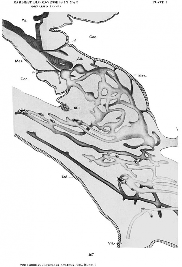
In the Minot embryo, by the graphic reconstruction method, it is easily seen that on the yolk—sac, between the entoderm and the mesoderm, there is a net of vascular tissue, one—layered and consisting of blood~islands, solid cords and angiocysts. This net is best developed, and the blood—islands in it are located exclusively, in the hemisphere of the yo1k—sac opposite the embryonic shield; this fact was pointed out by Minot in his article on the development of the blood in the Keibel and Mall “Human Embryology,” where it is spoken of as suggesting the area opaca and area pellucida of lower forms. But, although the vascular net is much more prominent near the distal pole of the yolk—sac, the net of slender solid cords can be traced, in the area pellucida, nearly to the embryonic shield. Before reaching the shield, the net in this embryo comes to an end, though apparently unconnected cords, resembling the angioblast cords, may be seen here and there running for only short distances, extending even into the bodystalk.
In the chorion and chorionic villi of the same embryo there are many of the “irregular spaces” described by other authors; they are cavities in the mesoderm, with distinct walls formed either by flattened or spindle—shaped cells, or merely by the clean cut edge of the loose mass of mesenchymal processes and fine fibrils in which they lie. In single sections the larger cavities may appear absolutely empty of any cellular content, but on reconstruction each cavity is always found to contain a shred of tissue, apparently floating in it. Not infrequently these shreds enclose small vacuoles, or may even open out into angiocysts, with a single layer of cells forming the wall. They occupy only a small part of the mesodermal cavities, as a rule, and seem to be loose in them, like a thread run through a pipe; often they lie close against the walls of the cavities. The impression received from the study of these shreds is that of an endothelium shrunken away from the rest of the Wall of a vessel. By making graphic reconstructions of these inner shreds of tissue, I found that here too the pattern obtained is that of a net, differing however from that on the yolk-sac by extending in three planes, or being in several layers. The reconstructions are not complete, in that many of the smaller branches were not traced to their termination, since it was chiefly desired to emphasize the net character of these cords.
This characteristic arrangement of these cords and vesicles, their general resemblance to other early endothelium, and other facts to be brought out later convince me that we are here dealing with the angioblast, or, perhaps better expressed. the endothelium of future blood-vessels.
The net in the chorion can be traced into many of the chorionic villi, and into the body-stalk as shown in figure 1; but in the bodystalk there is a great difference in the character of the elements composing it. The shreds or cords lying in relatively large mesodermal clefts open out into large endothelial angiocysts, from which cords again lead directly into large spaces in the mesoderm, without separate endothelial lining, or else end abruptly. I shall speak later of the histological appearances of these spaces, and for the present call them the “unlined spaces.” Other cords, not connected with the net, can be traced further toward the embryonic shield, along the allantois, but there is no direct connection with the yolk-sac net. The unlined spaces and the terminal cords are always near the surface of the body—stalk.
Grosser first called special attention to the epithelial layer of mesodermal cells, the mesothelium, which forms the coelomic surface of the yolk-sac and of the body-stalk in his 1 mm. embryo, ending abruptly at the junction of body—stalk and chorion. Moreover, he pointed out that this mesothelium, instead of forming a smooth surface, dipped in irregularly, giving in sections the appearance of festoons. This disposition of the mesothelium is found in all the young human embryos which I have studied, except the Bryce-Teacher ovum, in which there is as yet no coelomic cavity. A model of the body—stalk of the Minot embryo, in which the mesenchymal core is left out, leaving the mesothelium as a‘ sheet of Wax, enclosing the vascular net, shows the outer or coelomic surface formed of rounded ridges and irregular mounds, between which are deep cleftsc or depressions; on the inner surface of the mesothelium these clefts are seen as sharp ridges or pointed funnel-shaped ingrowths. The model shows clearly that these ridges and ingrowths touch here and there, and are apparently continuous with, either the angioblasti cords or the unlined spaces of the vascular net.
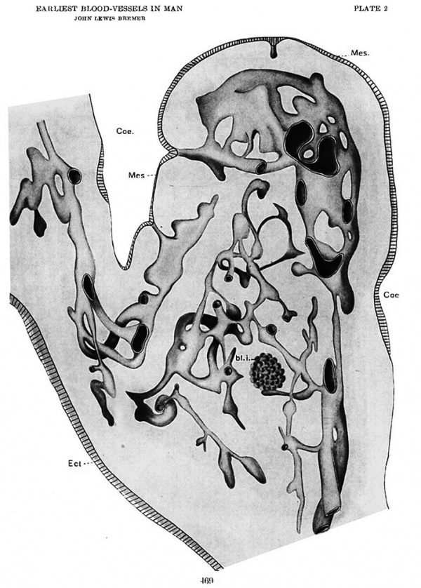
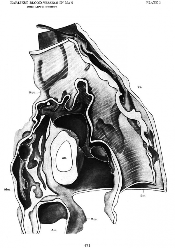
Through the great generosity of Professor Grosser I was enabled to repeat my reconstructions from his embryo, which‘ is faultlessly preserved, stained, and sectioned. The results correspond with the findings in the Minot embryo-, except that in Grosser’s embryo it is possible to trace the net from the chorion through the body stalk further toward the yolk-sac, and to follow one cord in its tortuous course to its junction with the yolk—sac net. Other cords from each net are apparently reaching out toward the other, some having nearly spanned the intervening space (f gs. 2 and 3). There are no cords unconnected with either net, such as are found in the Minot embryo. The end of the chorionic net nearest to the yolk-sac is composed of cords and unlined spaces, with no endothelial angiocysts.
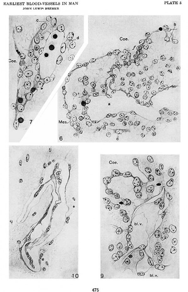
A careful study of the unlined spaces in this embryo leads to more convincing proof that they and the mesothelial ingrowths are actually connected. In the Minot embryo such connections can be made out only by reconstructions, whereas in the Grosser embryo the plane of ‘section has fortunately, in several instances, shown the whole connection in a single section, as shown in figures 6 and 7. Cells resembling young blood corpuscles may be very infrequently found in the unlined spaces, and even in the mesothelial ingrowths.
The blood—island described by Grosser as lying in the mesenchyma of the body-stalk of his embryo, is found to be connected by a protoplasmic cord, resembling an angioblast cord, with the vascular net (fig. 42). In the Minot embryo a few smaller but otherwise similar groups of cells are found, one of which is shown (fig. 1) connected directly with an inpocketing of the mesothelium. In both embryos the cells of the islands are rather small, rounded, with little protoplasm, resembling in these respects the cells of the young islands of the yolk-sac. In the body—sta1k of the Dandy embryo though it is much older than the two under consideration, I have found similar islands, one directly connected with an inpocketing of the mesothelium but not with the blood vessels, the other smaller one, of about 20;; in diameter, with no present connection with either blood-vessel or mesothelium.
In the embryo described by Professor Debeyre which he also most kindly allowed me to examine, the relations of mesothelium, unlined spaces, cords, angiocysts, and blood island appear to be the same; but no reconstructions were made.
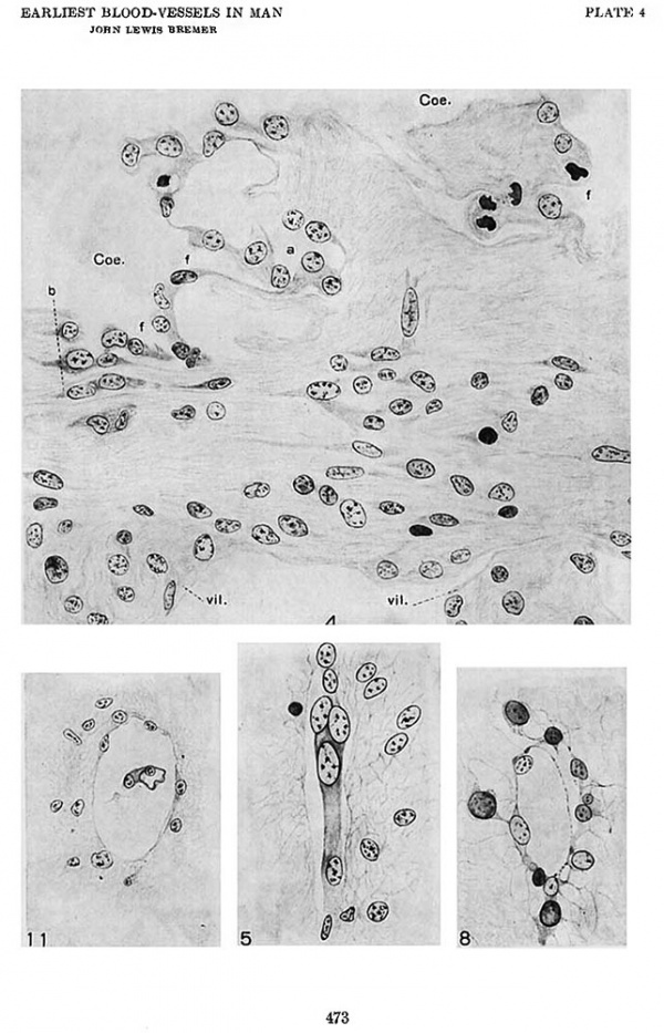
Herzog, it will be remembered, described in his embryo certain rings and small groups of cells, lying in the coelom at the edge of the body—stalk, which he considered the anlages of the yolk-sac Vessels. This valuable Very young embryo has been kindly given to the Harvard Embryological Collection, so that I have had the opportunity to study it carefully; and in spite of fact that the unfortunate breakage of many sections makes complete reconstructions impossible, enough can be seen to give a sure basis to the following account of its vascular anlages. After the study of the Minot and Grosser embryos it is not difficult to recognize that the groups and rings of cells in question are in fact tangential sections of the mounds of the mesothelial sheet covering the bodystalk; the lumen of the rings is the very loose mesenchymal core of the stalk. As is seen in the drawing (fig. 4) the mesothelial covering is not complete in this embryo, but leaves large areas of the surface uncovered, where the processes of the mesenchymal cells of the core, with numerous intervening fibrils, form the only border between the body-stalk and the coelom, as is the case along the inner border of the chorion proper. Where the mesothelial cells are present they occasionally project into the core of the bodystalk, lining a funnel-shaped diverticulum of the coelom, as is seen also in the older embryos. Mitotic figures are frequent at such points. The inner end of such projections are continued, usually in a curved direction, as irregular hollow spaces (ii g. 4, a), as cords or as small groups of cells, without lumen. One of these cords, three or four cells long, runs in the chorion parallel to its inner border. It seems very probable that from the.ceIl groups other cords also run in the chorion, but the destruction of certain sections makes positive proof of this impossible. One especially large and well defined group of cells lies at the base of the body—stalk, in the mesenchyma near the coelomic border, and directly over it is found a mesothelial sheet and inpocketing; but here again the condition of the sections makes it impossible to affirm that the two structures are actually continuous. In the chorion there are none of the large irregular spaces seen in older specimens; but, extending from the base of the body-stalk for a considerable distance, perhaps a quarter of the way around the chorion, are cords of cells Which, on reconstruction, are found to form an irregular net, in one plane, parallel to the surface of the coelom. This net shows no trace of angiocysts, nor are there even vacuoles in the cell protoplasm. The cells forming it are broader, or thicker, than the surrounding mesenchymal cells, many of which are spindle-shaped, and the cord cells usually lie in well defined, though small, cavities among the mesodermal fibrils and cell processes (fig. 5).
The yolk-sac of the Herzog embryo is covered by a mesothelium, apparently originally complete, though broken now in many places but this mesothelium lies everywhere close against the enclosed entoderm, and shows no signs of the funnel-shaped ingrowths such as are found around the body-stalk. In Jung’s paper, however, (his F g. 17) the mesothelium of the yolk—sac is shown as quite far separated from the entoderm, and in at least two places in the one drawing (the only section of the embryo proper figured by him) the mesothelium of the distal pole is arranged in what appear to be typical funnels.
In the Bryce—Teacher ovum, which was also most generously placed at my disposal for examination, the mesodermal cells are often spindle~shaped, and may be arranged in chains of two or three cells, followable from one section to another. But on reconstruction these cells do not form a net, except by the very finest protoplasmic processes, as in ordinary mesenchyma. A coelom is absent, and therefore there is no mesothelial surface. Though the chains of cells look somewhat like angioblast cords, I am satisfied that they are only portions of the general mesenchyma, especially since similar chains of spindle—shaped cells, followable for only short distances, and not connected with the vascular net, are to be found in the mesenchyma of older embryos, even at the time‘ when the blood—vessels are well established. Much larger, more distinct cords of cells in the mesoderm of this ovum are found to be processes of the surface ectoderm, of which a few cords traverse the mesoderm of the future chorion. Similar, but hollow, “chorionic canals” are described by Grosser and by F. T. Lewis[9] in older embryos. From my study of this ovum I should say that no vascular anlages exist at this stage.
I have mentioned the “unlined spaces” which seem to form a part of the vascular net, and wish now to describe them and the other portions of the net in some histological detail. In the Herzog embryo (fig. 4) a long funnel-shaped diverticulum of the coelom, bounded by a definite mesothelial layer, is seen to expand at its distal end (a). The two walls approach each other in the middle portion of the diverticulum, and are either in contact or definitely fused, thus cutting ofl' a distal cavity. If the fusion of the walls had continued further toward the main coelom, the distal cavity would appear as an irregular space, deep in the mesenchymal core of the body-stalk, but connected with the mesothelial surface layer by a cord of mesothelial cells. Moreover, the cavity would be a portion of the coelom, and its bounding walls would be also mesothelium. If we turn now to the Grosser embryo, we see another cavity (fig. 6, a) ; the walls are histologically similar to the mesothelium (mes.) covering the body—stalk at this point, and are connected with the surface layer (at the top of the drawing) by a cord of the same type of cells. The funnel-shaped mouth of the original diverticulum is still clearly seen. Both on the coelomic surface and bounding the cavity, the protoplasm of this layer forms a narrow but definite sheet, and is connected by numerous processes with the underlying mesenchyma. The nuclei are broader than the sheet of protoplasm, and project now toward the cavity, now toward the mesenchyma. The mesothelium of the surface, the mesothelial cord, the walls of the enclosed cavity, and the surrounding mesenchyma. all form a syncytium, as no cell walls are present. The shape of this-cavity as traced through the sections and the fact that in other sections there are other mesothelial cords connecting it with the surface make it probable that several smaller cavities have coalesced to form this one. A much smaller cavity is seen in the same drawing at b, near the surface.
Another similar cavity, also from the Grosser embryo (fig. 7), shows the same characteristics as the last described, except that the funnel-shaped opening into the coelom has apparently become obliterated by a more complete fusion of its walls, so that the mesothelial cord now springs from the under side of a smooth portion of the surface layer. Here again the walls of the cavity are histologically gimilar to the boundary of the eoelom and to the mesothelial cord. In this case the walls are continued in two directions by protoplasmic strands (c and cl) broader than the ordinary rnesenchymal processes, with which they connect, and of the same character as the strand joining the walls of the cavity with surface mesothelium. One of these (c) leads to a second funnel on the surface, and merely proves the coalescence of two separate ingrowths; the other (cl) ends blindly, as is shown in the drawing, and indicates either that the distal, as well as the proximal end of the original coelomic diverticulum may be obliterated by fusion, or (which seems to me more probable) that the walls of the diverticulum have the power of further growth. In figure 6 another similar strand is seen at c, which in this section appears isolated, but by reconstruction is found to connect two adjacent cavities. The relation between these unlined spaces, which from my drawings I can only consider as isolated portions of the coelom, and the angiocysts which have a definite endothelial lining and an extraintimal space, perhaps due to shrinkage, is indicated in the next two drawings. One, from the body—stalk of Grosser’s embryo, (fi g. 8) shows the typical mesothelial wall on one side of the cavity, and on the other an apparent delamination of an inner layer, continuous at either end with the mesothelial wall, but separated as a whole by an extra-intimal space from the underlying layer, which is still an integral part of the surrounding mesenchyma. The other drawing (f g. 9) is from the Minot embryo, near the wall of the coelom at the side of the body—stalk. Two funnel-shaped diverticula from the coelom lead toward the inner cavity, though the mesothelial cords are not so distinct as in the former cases. The wall of the cavity is mesothlelial at the lower left-hand corner, but is continued as an inner lining, with an extra-intimal space. This inner lining, in the form of an extremely thin sheet of tissue containing scattered nuclei, running in part obliquely through the section, in part directly away from the eye of the observer, sends out, in three directions, processes which connect with the inner lining of other similar cavities; one such connection is shown in the drawing, the others are continued in adjoining sections. We are dealing here, undoubtedly, with endothelium, and the processes are typical angioblast cords. The cords run usually, if not always, in well defined spaces in the mesenchyma.
The mesothelial cords, as well as the processes of the surrounding mesenchymal cells, are left, as it were, attached to the outer wall of the cavity, while the endothelium lies free within. Such mesothelial cords are found in much older embryos, as for instance in that described by Dandy (of seven segments) and in one of about the same age kindly lent me by Prof. R. Meyer of Berlin. They lead from the outer walls of the now well defined umbilical vessels to funnel-shaped irregularities of the coelom wall, where they are continuous with the surface mesothelium, frequently anastomosing with each other. Since they seem involved in the process of haemopoesis, I shall mention them again later.
In addition to the method just described, whereby endothelium is derived by delamination from the mesothelium of isolated portions of the coelom, a more direct method seems to be shown in the preparations studied. It will be remembered that in the reconstructions of the Minot embryo (and the same is true of those of the Grosser embryo) the angioblast ‘cords of the net could be frequently traced to ingrowths from the coelomic mesothelium without the intervention of the unlined spaces. Isolated portions of such cords, in advance of the -net in the Minot embryo (fig. 1) are also thus connected. Again, in the Herzog embryo, one of the funnel-shaped-ingrowths is continuous, as seen by reconstruction, with a short cord (E g. 4, b) which it is impossible to follow far, but which is similar to those forming the net in the chorion. These cords are characterized by rather darkly staining protoplasm, and by the absence of protoplasmic processes connecting them with the surrounding mesenchyma, from which they are usually slightly separated, leaving an extra—intimal space (fig. 5). Their resemblance to the endothelial cords which form the links between angiocysts in all the vascular nets studied makes me believe that they also are true endothelium, and that thus endothelium may arise directly as an extension of the mesothelial cords, without the process of delamination. Any future cavity in these cords would be potentially a part of the coelom.
If these interpretations of the sections are correct, true endothelium may arise in two ways from the mesothelium. Certain appearances make it at least probable that blood corpuscles may also be a product of the same tissue. In the yolk-sac blood-islands it has long been agreed that both endothelium and corpuscles come from the same anlages. In the body-stalk net of the Minot embryo, which is not connected with the yolk-sac net, there are a very few blood-corpuscles, or at least free cells within endothelial cavities. In the Grosser embryo, in which the connection of the two nets is apparently solid, there are also a very few corpuscles. In both of these embryos the yolk-sac corpuscles are limited to the distal pole; it seems certain, therefore, that the few blood—corpuscles in the body-stalk vessels must have developed in the net itself. The blood—islands. already mentioned in the body-stalks of these two embryos are either not connected with the other vessels, or are so connected only by apparently solid strands (figs. 1 and 2); they will probably later supply their quota of corpuscles, but do not seem to account for the few already present.
In the embryo of Professor R. Meyer, already described as showing mesothelial cords still connecting the blood-vessels with the coelomic wall, the blood-corpuscles are differentially stained, and easily distinguishable from other cells. They are found not only in the blood-vessels, but also in the mesothelial cords, of which they seem to form a part. Similarly in the drawing of the Grosser embryo (fig. 7) there are, in -and connected with the mesothelium, cells whose nuclei resemble those of the three cells floating free in the cavity. Though these cells are not of the type of corpuscle which one would expect at such an early age, cells of the same character are found occasionally in the. yolk~sac vessels of both the Grosser and the Minot embryos, and from their position I consider it probable that they are blood-corpuscles. We should, then, credit this mesothelium with the additional power of forming occasional corpuscles without the mediation of blood-islands.
Briefly summarized, my observations point to ingrowths of the mesothelial layer covering the yolk-sac and body-stalk as the anlages of the blood-vessel endothelium and, to a lesser extent, of the blood-corpuscles. The anlages, though limited to certain areas commensurate with the extent of the mesothelial layer, are multiple and form a net by the growth and coalescence of the separate units. A further extension, accompanied by the incorporation of other units, effects the union of the body-stalk net with that on the yolk-sac. On the other hand, the net in the chorion and chorionic villi seems to me to be formed by direct extension from the body-stalk, without the addition of new components, and to be the result of a centrifugal growth of endothelial sprouts, in the form of angioblast cords, with here and there expanded angiocysts, which advance through the mesenchyma. This would correspond with the fact that no surely isolated endothelial cords have been found in the chorion, that in the Herzog embryo the net extends only part way around the chorion and is centered at the base of the body-stalk, and that there is in the embryos studied no mesothelium on the inner surface of the chorion proper, and therefore‘ no possibility of new anlages there. The chorionic mesothelium is developed later, but only after the vessels are well established.
That the angioblast cords and angiocysts are direct sprouts from the endothelium and not mesenchymal cells metamorphosed in situ and added to the growing tip is shown (aside from the recent work of Clark[10] and others on endothelial growth) by the presence of the clear-cut extra-intimal space (figs. 10 and 11) and by the fact that there are no protoplasmic connections between endothelium and mesenchyma, even in the most distal vessels. The irregularities occasionally found in the outlines of the endothelium seem to me to point to its contraction or perhaps to amoeboid movements incident to the formation of new sprouts. That the extra-intimal space may be an artefact, that the so—called cords may be in reality collapsed tubes, is, to my mind, immaterial, and awaits proof by injection methods; the sharp boundary between the endothelium and the mesenchyma through which it runs points to a difference in origin between the two tissues.
We have not yet attained a sure histological basis for differentiating endothelium in ordinary specimens, nor is it perhaps to be expected in tissues as young as those under discussion. In a recent paper Clark[11] described certain differential characteristics of endothelial nuclei, after special fixation and stains, but this is in chicks of a relatively older stage than the present human material, and I could find no trace of such differences in younger material and with the common stains used. In the Grosser embryo, as shown in many of the drawings, endothelium and mesothelium are both often marked by the presence of very fine intra-cellular fibrils, absent in the mesenchymal cells; these fibrils are not found in the Minot embryo, nor in any of the others studied, owing probably to differences in the fixing fluid employed. They cannot, therefore, be used as a general distinguishing sign.
The coincidence of the views forced upon me by my observations with the now ancient theory of His,[12] Butschli,[13] and others, that blood-vessels are in some way related to the coelom, is apparent, and the significance of this when correlated with the facts known of the inter-relation of the blood-vascular system and the coelom in certain invertebrates, must strike anyone who is interested in phylogeny.
This has been briefly noted by Hungtington[14] who found, in a cat embryo of 10 mm., a “clearly limited and well defined funnel shaped stoma, occupying the dorsal extremity of the coelomic cleft and apparently opening directly into the spaces of the early lymphatic plexus” (p. 26), without, however, attaching more than a “suggestive” importance to the fact. It is also interesting to note that the origin of endothelium from the mesothelium adds to the already great number of structures derived by ingrowths from this source, among which may be mentioned the tubules of the pronephros (indirectly also those of the mesonephros), the sex cords, and the cortex of the suprarenal gland, according to the later views.[15]
The origin of the chorionic vessels from the mesothelium of the body-stalk explains the fact, mentioned by Knoop[16] in anomalies of the amnion, and by Bauereisen[17] in haematomoles, that vessels can exist in the chorion “without the help of the umbilical Vessels.”
Let us now turn to a discussion of the papers before referred to, in which the authors describe the results of experiments, on chick or other bird embryos, undertaken to prove the origin in situ of intra—embryonic blood—vessels, as opposed to the extension or growth of such vessels from some extra-embryonic source, by destroying or cutting off the whole or part of the area opaca of one side. Vessels on the injured side are obtained in many cases by all these investigators, after a further incubation of the operated embryos} and these vessels are generally in the position of aorta, heart, Vitelline vein, and even other Vessels of normal embryos. Where only part of the vascular system exists on the injured side, ‘it appears to be always that nearest the mid-line; i.e., the aorta may be present without the heart, or the aorta and heart without the other vessels. Not infrequently the vessels are abnormally large, or the lateral vessels may be only roughly in the normal position. Graper[18] and Hahn,[19] though the results of each were known to the other before publication, come to different conclusions as to the origin of the vessels ; the first maintaining from his experiments, an entodermal, the other from_ his experiments a mesodermal derivation. Miller[20] is chiefly interested in the negative proof that the intraembryonic vessels do not reach their ultimate destination by direct ingrowths of endothelium from the lateral area, as was maintained in my former paper.
From the differences in the conclusions reached by two of these authors it seems certain that more work should be done along these lines before a consensus of opinion can be expected. I wish to point out a few possibilities which should, I think, be considered in any such future work.
As was shown in the figures of my reconstructions of rabbit embryos, the vascular net has an irregular mesial border, certain strands lying further toward the midline than the position of the future aorta. Though in the younger embryos the extension of these strands across the median line of the embryo proper to ‘form a net on the opposite side is rendered impossible by the close approximation of the medullary groove, notochord, and entoderm, yet long before the stage figured in many instances cited by these authors the mesoderm has grown across the median line, and might afford a pathway for endothelial sprouts from side to side. Another and earlier pathway is at the posterior end of the embryo, behind the primitive streak, where the mesoderm very early extends across the median line. The angioblast cords, by which connections from recognizable blood-vessels to apparently isolated angiocysts can be traced (if we accept, for the moment, and for the purpose of argument, the extension theory) are delicate strands, easily overlooked, and moreover may last only a few hours, if the mechanical conditions are not favorable to their continued development into vessels (cf. figs. 1 and 3, loc. cit.). It is not to be expected, therefore, that anything short of a very complete series of such operated embryos, fixed at progressively longer intervals of incubation after operation, can settle whether or not there is any extension from the opposite side.
I think it well at this point to define more accurately what I mean by angioblast cords, especially since I believe that their recognition may perhaps help to explain the frequently described endothelial spaces unconnected with any injectable vessels. The angioblast cords are apparently solid cords of cells, connected end to end or in small groups, running between the processes of the surrounding mesenchymal cells, when these are present, often touching them, without however actually fusing with them. The diameter of the cords is never as small as that of the mesenchymal processes, though it is often less than that of the cord nuclei. The cords tend to form nets by anastomosis of larger mesh than the mesenchymal net, and angiocysts. by vacuolization wherever space is given. They are usually sharply defined from the surrounding tissue, and may show an extra—intimal space. They must necessarily be extremely hard to recognize in dense mesenchyma, though easy to trace in perfectly prepared series of looser tissue.
A second possibility to be considered in attempting to explain, on the basis of the specificity of endothelium, the presence of vessels on the injured side of the operated embryos is advanced in this present paper.‘ If the earliest human vessels arise from the mesothelium lining a portion of the extra—embryonic coelom, by multiple anlages which later fuse; and if the intra-embryonic coelom, developed later, is also lined by mesothelium, the opportunity is perhaps offered for similar, but later, anlages for intraembryonic vessels. The instance cited by Huntington ‘of a coelomic opening into the spaces of the early lymphatic plexus should be borne in mine. I am well aware that the yolk—sac blood-islands are not proven to arise from mesothelial anlages, that in avian embryos they are even said to be present before the formation of the coelom in the area Vasculosa, a fact that would point to some tissue other than mesothelium (perhaps a premesothelial stage of mesoderm) as that from which endothelium is derived; yet certain connections between the vessels of the operated embryos above referred to and the mesothelial wall of the coelom, shown in many of the drawings and seen by me in one of Miller’s preparations, seem to me to make this possibility at least worthy of consideration in future study. It is easily conceivable that such separate anlages might arise under abnormal conditions in positions where they are normally absent; or on the other hand it may be that multiple anlages of intra—embryonic vessels in close relation to the coelom are normal, and that the net figured by me in this position is the result of their‘ confluence. Yet the finding of no separate angioblast cords in advance of the general net in this specimen, especially at the younger caudal end, would militate against this latter proposition. It is also possible that embryos of such different types as chick and rabbit or man may show differences in details of the vascular development.
Once given an endothelial net in the general area of the future vessels, mechanical forces would, as pointed out in the paper so often referred to, locate the portions of that net which are to remain and form aorta, heart, etc., and the portions which are to disappear because unfavorably placed. It is probable that all the variations in size and shape of heart and aorta, and even in position of the more lateral vessels, which are not infrequently shown in the drawings of the operated embryos, are due to changes in the normal tension of the germ layers, consequent on the injury. That these changes must be great is shown in gross by the bending of the whole body of such embryos away from the injured side. A study of these embryos from this point of view would lead to interesting results in the field of developmental mechanics.
Conclusions
In human embryos the earliest blood-vessels arise separately in the yolk—sac and in the body-stalk, by multiple anlages.
The anlages in the body-stalk (and perhaps also in the yolk—sac (cf. Jung’s figure 17) are funnel-shaped ingrowths of the surface mesothelium, which is present as a definite layer only on the two areas mentioned. By partial fusion of the walls of an ingrowth a portion of the coelom, still bordered by mesothelium, may be cut off as a separate cavity, lying deep within the substance of the body-stalk.
The endothelium seems to arise either (a) by delamination from the walls of such a detached portion of the coelom, or (b) by direct extension, in the form of an angioblast cord, from the mesothelial ingrowth. From the endothelium, by whichever method developed, further extension is by means of the angioblast cords, which grow apparently through the surrounding mesoderm.
True blood-islands may occasionally arise by the multiplication of the cells of the mesothelial ingrowths, or scattered blood corpuscles may arise singly within these ingrowths.
Extension within the limit of the areas covered by the mesothelium is achieved by confluence of the detached portions of the coelom, or union of the cords; the result is a net comprising the various vascular units. Extension into the chorion, where the mesothelial layer is absent in the early stages, appears to be by direct centrifugal growth of the angioblast cords, Without the addition of new elements from the surrounding mesenchyma.
The possibility that similar, but later, ingrowths from the mesothelium of the intra-embryonic coelom may give rise to intra-embryonic vessels, should be borne in mind in the study of such vessels, Whether haemal or lymphatic.
References
- ↑ Bremer JL. The development of the aorta and aortic arches in rabbits. (1912) Amer. J Anat. 13(2): 3-128.
- ↑ Grosser, O. Anat. Hefte. Bd. 47, p. 649, 1913.
- ↑ Debeyre, A. Journal de l’Anat. et de la Physiologie. Vol. 48, p. 448, 1912.
- ↑ Jung. Miinchen. med. Wochenschr. Jahrg. 54, p. 1343, 1907.
- ↑ Herzog, M. Am. Jour. Anat. Vol. 9, p. 361, 1909.
- ↑ Frassi, L. Arch. mikr. Anat. Bd. 70, p. 492, and Bd. 71, p. 667, 1907—08.
- ↑ Dandy WE. A human embryo with seven pairs of somites measuring about 2 mm in length. (1910) Amer. J Anat. 10: 85-109.
- ↑ Bryce, T. H. and Teacher, J. H. Memoir, 1908.
- ↑ Lewis, F. T. Communication to the 30th Session of the Am. Ass. Anat., 1913.
- ↑ Clark, E. R. Am. Journ. Anat. Vol. 13, p. 351, 1912.
- ↑ Clark, E. R. Anat. Record. Vol. 8, p. 81, 1914.
- ↑ His, W. Abhandl. math. phys. Classe K. Sachs. Ges. Wiss. Vol. 26, p. 173, 1900.
- ↑ Biitschli, O. Morph. Jahrb. Bd. 8, p. 474, 1883.
- ↑ Huntington, G. S. Memoirs Wistar Institute, No. 1, 1911.
- ↑ Goormaghtigh, N. Bull. Soc. Med. de Gand. Vol. 5, p. 24, 1914.
- ↑ Knoop, H. Beitr. Geburtsh. Gyniik. Bd. 7, p. 284, 1903.
- ↑ Bauereisen, quoted from Jagerroos, B. H. Archiv f. mikr. Anat. Bd.82, Abt. '1, p. 271, 1913.
- ↑ Graper, L. Arch. f. Entw. Mech. Bd. 24, p. 375, 1907.
- ↑ Hahn, H. Arch. f. Entw. Mech. Bd. 27, p. 337. 1909.
- ↑ Miller, A. M. Anat. Record. Vol.8, p. 91, 1914.
Plates
| Historic Disclaimer - information about historic embryology pages |
|---|
| Pages where the terms "Historic" (textbooks, papers, people, recommendations) appear on this site, and sections within pages where this disclaimer appears, indicate that the content and scientific understanding are specific to the time of publication. This means that while some scientific descriptions are still accurate, the terminology and interpretation of the developmental mechanisms reflect the understanding at the time of original publication and those of the preceding periods, these terms, interpretations and recommendations may not reflect our current scientific understanding. (More? Embryology History | Historic Embryology Papers) |
Plate 1
Minot embryo, H.E.C. 03 . 825. Reconstruction of vessels in the body-stalk and chorion; connections with the mesothelium. All., allantois; bl.i.. blood-island; Coe., coelom; Ect., ectoderm of chorion; f. , funnel-shaped connections, with unconnected cords; Mes., mesothelium; Vd., villus; Ys., yolk-sac cavity. X 175.
Plate 2
Grosser embryo, slide 3, row 5. Reconstruction of vessels in the body-stalk and chorion. Lettering same as in figure 1. X 275.
Plate 3
Grosser embryo, slide 3, row 3. Junction of body-stalk and yolk-sac, and connection of the two vascular nets, along the side of the allantois, between the mesoderm and entoderm. All., cavity of allantois, looking into yolk-sac; Am., cavity of amnion, which at one point is fused with the allantois;Ent., sheet of entoderm forming wall of yolk-sac and continued as allantois wall; Mes., sheet of mesothelium continued from yolk-sac to body-stalk; Ys., cavity of yolk-sac. X 400.
Plate 4
4 Herzog embryo, slide M, sect. 147. Chorion and base of body-stalk, to show irregularity of the coelomic surface, the partial mesothelial layer, and the funnel-shaped ingrowths cf.) The chorionic ectoderm has shrunken away, leaving naked the chorionic mesoderm. at the bottom of the drawing, znd the stumps of two villi (zd.).C'oe.,coelom;a, unlined space;b, angioblast cord. X circa800.
5 Herzog embryo, slide RI, sect. 143. Angioblast cord in chorion, part of a net. Notice t h e clear-cut extra-intimal space. X circa 800.
8 Grosser embryo, slide 3, row 8, sect. 3. Vessel from the body-stalk to show delamination of endothelium and formation of extra-intimal space. X circa 800.
11 Minot embryo, H.E.C. no. 825, sect. 27. Cross section of a strand of the net in the chorion to show a small angiocyst. X circa 540.
Plate 5
6 Grosser cmbryo, slide 3, row 5, sect. 3. From the edge of the body-stalk, to show surface mesothelium (Mes.) and mesothelial cord, leading from funnel (f.) to unlined space (a). Coe., coelom; h, smaller unlined space;c, endothelial cord. X circa 580.
7 Grosser embryo, slide 3, row 4, scct. 5. From the edge of the body-stalk, to show mesothelial cord leading to unlined space, containing corpuscles. c and d, mesothelial cords (see text); Coe., coelom. X circa 800.
9 Minot embryo, H.E.C. no. 825, sect. 25. Vesscl from the body-stalk, to show delamination of endothelium, and its extension as cords. Further descrip- tion in text. Coc., coelom; ~Z. U. , blood-vessel. X circa 800.
10 Minot embryo, H.E.C. no. 825, sect. 24. Angioblast net in the chorion, to show the irregular contour of the cord, suggesting amoeboid movements, and the extra-intimnl space even around the young branch. X circa 540.
Cite this page: Hill, M.A. (2024, April 27) Embryology Paper - The Earliest Blood-Vessels in Man. Retrieved from https://embryology.med.unsw.edu.au/embryology/index.php/Paper_-_The_Earliest_Blood-Vessels_in_Man
- © Dr Mark Hill 2024, UNSW Embryology ISBN: 978 0 7334 2609 4 - UNSW CRICOS Provider Code No. 00098G


