Paper - Testes descent 1909 - 1
| Embryology - 26 Jul 2026 |
|---|
| Google Translate - select your language from the list shown below (this will open a new external page) |
|
العربية | català | 中文 | 中國傳統的 | français | Deutsche | עִברִית | हिंदी | bahasa Indonesia | italiano | 日本語 | 한국어 | မြန်မာ | Pilipino | Polskie | português | ਪੰਜਾਬੀ ਦੇ | Română | русский | Español | Swahili | Svensk | ไทย | Türkçe | اردو | ייִדיש | Tiếng Việt These external translations are automated and may not be accurate. (More? About Translations) |
Hart DB. The nature and cause of the physiological descent of the testes. (1909) J Anat Physiol. 43(3): 244-65. PMID 17232805
| Historic Disclaimer - information about historic embryology pages |
|---|
 |
The Nature and Cause of the Physiological Descent of the Testes
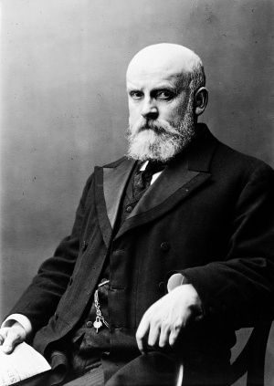
By D. Berry Hart, M.D., Etc.,
Lecturer on Midwifery, Surgeons’ Hall, Edinburgh; Hon. Fellow, American Gynaecological Society ; Carnegie Research Fellow.
This inquiry was carried out at the Laboratory of the Royal College of Physicians Edinburgh. (1909)
Part I. Descent in Marsupials
“At this time of life the testis is connected in a very particular manner with the parietes of the abdomen, at that place Where in adult bodies the spermatic vessels pass out, and likewise with the scrotum. This connection is by means of a substance which runs down from the lower end of the testis to the scrotum, and which at present I shall call the ligament, or gubérnaculum testis, because it connects the testis with the scrotum and directs its course in its descent ” (p. 6).
“ It is plain from this description, that the cavity of the bag or of the elongation of the peritoneum which contains the testis in the scrotum, must at first communicate with the general cavity of the abdomen by an aperture at the inside of the groin” (p. 11).
“When the testis is descending and when it has even passed into the scrotum, it is still covered by the peritoneum exactly in the same manner as when it was in the abdomen” (p. 10).—John Hunter, “A Description of the Situation of the Testis in the Foetus, with its Descent into the Scrotum,” in Observations on Certain Parts of the Animal (Economy, London, 1786.
“ The parts in the female appropriated for the purpose of supplying the young are variously placed in different animals. In the horse, black cattle, sheep, and other graminivorous animals, their situation is between the hind legs, which is also the place allotted for the testicles of the male of this tribe ; (and probably of all those in which they come out of the cavity of the belly) therefore in the hermaphrodite which has both these parts, the testicles are in some degree obliged to come down into the udder, which does not receive them so readily as the scrotum.”-John Hunter, “Account of the Free Martin,” in p. 45, op. cit.
In a previous communication I described a central canal in the ovary of the rat-kangaroo, apparently derived from the Mullerian duct. The question arose as to whether this was exceptional or normal in marsupials, and therefore, at Dr James Ritchie’s suggestion, and through his kind services, I obtained from Professor Wilson of Sydney the pelvic ends of five kangaroo embryos in good preservation. Four of these were Macropus rw/icollis; the fifth and largest, Dasg/urns viverrinus. After suitable preparation, they were cut in transverse serial sections and stained with logwood and eosin. In this way about three thousand sections were obtained. I now found that two were female and three male. This seemed at first a disappointment; but, on examination, the male embryos were found to be of great interest and novelty in many points, so that the results of their examination and of an extended investigation into the whole question form the subject of the present paper.
The results of the examination of the female specimens, so far as the development of the mammary pouch and round ligaments is concerned, will be found in another paper; but the conditions in the two sexes are intimately connected with one another, and must be read together to get a complete view of the subject. I found no central canal in the ovary.
In two of the male specimens (Mctcropus ruficollis) the Wolflian bodies were present with the developing testes on their inner and upper surfaces ; the inguinal canal, gubernaculum, and cremaster had developed, and the scrotum was developing; in a third, more advanced, the Wolfiian bodies had disappeared, the scrotum had developed and separated, and the testes were in the inguinal canal. One special point of value in the last specimen was that, although the testes were in the inguinal canal, its size was such that serial sections could be conveniently made. In the human male foetus at this stage the specimens are too large for this being done.
I shall therefore take up I. The development of the scrotum, inguinal canal, and gubernaculum prior to the descent of the testes. II. The condition of the scrotum and inguinal canal after the testes were in the inguinal canal. ' III. The mechanism of descent of the testes as shown in the marsupial specimens. IV. The descent of the testes in the human foetus. V. The phylogeny of descent of the testes and of the parts involved in it.
I. The Development of the Scrotum, Inguinal Canal, and Gubernaculum prior to the Desoent of the Testes
This was well shown in two specimens, but I describe the conditions in one only: the other corroborated these. The sections are first considered from above down, and begin a little above the suprapubic region.
Development of the Scrotum. Structure of the Abdominal Wall on Tr. Section. — The epidermis of the anterior abdominal wall in the suprapubic region has two ridges, one on each side of the middle line, the depression between the two lying opposite the linea alba. The deep epidermal cells are well marked, Behind it lies a layer of fibrous connective tissue, and then comes a layer of denser connective tissue continuous on each side with lymph-sinuses to be presently described. This special layer has a homogeneous ground-substance interspersed with connective-tissue corpuscles and unstriped muscle. Next comes a layer of mesenchymatous tissue, and finally the peritoneum. Lower down on the abdominal surface the epidermic ridges are larger in section, and each has in it a circular space filled with connective tissue of a myxomatous nature surrounded by deep epidermal cells. These are evidently gsections of double solid cones in the epidermis. The sections of the cones below this lose their intermediate epidermal septum, and We get a larger hour-glass space. Then they lose their posterior epidermal Wall, and the core has in it a double fan-shaped expansion of unstriped muscle derived from the inguinal fold, and broader in front (gubernacular fibres). There are thus formed in the suprapubic epidermic ridges double cones, complete above, but with the internal and posterior boundary walls incomplete lower down. They are formed by the deep epidermal layer passing backwards and snaring in the connective tissue completely above, but less completely below, where the epidermal horns do not meet. These cones—best termed the scrotal cones——form the scrotum, into which the testicles ultimately pass. The fully formed scrotum is solid at first, but ultimately develops a cavity as the gubernaculum grows into it, as Klaatsch and Frankl and Katz figure in their specimens (figs. 1, 2, 3, and 4).
fiG. 1.—T.S. embryo, Macropus ruficollis ; Wolflian bodies and testes in abdomen. (§—.) 1, epidermal ridges; 2, lymph-sinuses; 3, pyramidalis muscle.
fiG. 2. —T.S. Macropus ruficollis embryo. (5f’—.) 1, phallus; 2, scrotal cone ; 3, lymph-sinus. Note median partition disappearing.
The marsupial scrotum has long been known to be prepenial, but the reason has never been fully explained. All writers, except Klaatsch, consider it homologous and analogous to the ordinary scrotum in higher mammals. If so, however, it has not only appeared prematurely in phylogeny, but should not be prepenial and suprapubic. The account I have given of its development explains its nature. It is the analogue of the female mammary pouch, and, although this may seem paradoxical, its homologue too. It develops like the mammary pouch, but the deep epidermic layer passes backwards in crescentic form, the double fold containing little more than the deep epidermic cells, as the cavity of the pouch is to be formed, not by the desquamating epidermis as in the mamma, but from the enclosed connective tissue. This development of the scrotum in the suprapubic region necessarily makes it prepenial, and the desquamation of the cells enclosed by the deep epidermic horns at a later stage renders the scrotum free and pendulous.
After making out the above developmental points in -regard to the scrotum, I was glad to find it confirmed by K1aatsch’s opinion, given in his paper. He there suggests that, from its position and relation to the gubernaculum—the analogue of the round ligaments—the scrotum must be the analogue ofthe mammary pouch. This question will be discussed, however, later on. I may mention’ here that it occurred to me that the adult scrotum might show a furrow in its mesial aspect as evidence of this development. The specimen Klaatsch figures does not show this, but it is well seen in the scrotum of Acrobata pygmaza figured by Katz. He describes this scrotum as heart‘-shaped, and states that he has seen it in young male specimens from Brazil, and in an adult specimen of the opossum (fig. 5).
fiG. 3. M0w’I’070’t6S rufiwllis. (-'39.)
fiG. 4.—— Gubernacular fibres in scrotum. (1%-‘-’.) 1, gubernacular tibr i r t ; 2 ' te - ' b d d’ - ' ' ' placed up and bfixszkn; lg,’ Ilia 31:0! delelb eI‘p’ill1ia‘tl'ii1isa1'iassii18g 1' epldermal ridge (3 m fig’ 3). back; 4, solid scrotum formed from scrotal cones.
In the male embryo where the testes were in the inguinal canal, the scrotum was separated from its epidermal bed and projected at right angles from the abdominal surface. It was still solid (fig. 15).
I may sum up this point here, then, by saying that the scrotum in marsupials is the analogue and homologue of the mammary pouch of the female in every respect, but the full proof of the latter is only given in the section on Phylogeny (V.).
The Development of the Inguinal Canal and Gubernaculum. —— The development of the inguinal canal and its contents, excluding the testes and epididymis in the male, is intimately bound up with that of the lymphsinuses in the groin, and of the gubernaculum, acting in association with the muscular and fascia] structures in the abdominal wall on which the inguinal fold, to be presently described, abuts. In both the specimens we are now considering, the testes were abdominal and developing on the Wolflian bodies, ’i.e. were abdominal and high up. This fact must be emphasised, as we shall see just now that the inguinal canal had developed
fiG. 5.—Scrotum in Acrobata (§‘—.) a., anus ; p., phallus; 9., scrotum ; rr., groove between original cones. (Katz.)
while the testes were in the abdomen, so that, broadly speaking, the inguinal canal, contents and lining, tunica vaginalis, the gubernaculum (at this special stage), and cremaster owe their presence to a penetrating growth of gubernacular fibres, peritoneum, and cremaster along a definite lymphatic tract while the testes are abdominal, and thus not to any mechanical dilating process connected with the descent of the testicles. To understand this we must consider the structure of the abdominal wall. The anterior abdominal wall of the embryos, viewed on transverse section, has the following. structure. first, on the surface, on each side of the middle line, is the epidermal layer with the two ridges already described, in which the scrotal cones are developed. Behind this, comes a thin layer of unstriped muscle, and then a thicker layer of connective tissue of a mesenchymatous nature, but with fibrous cells condensing it. Next this, lies a deeply stained band of connective tissue joining the inner ends of the lymph-sinuses; then more mesenchymatous tissue; and lastly the pyramidales, recti, and transversales muscles, the external oblique and peritoneum, the last closely bound up with a fascia into which in the flanks the transversalis radiates. This fascia forms the transversalis fascia; the cord-covering known as the cremasteric fascia is derived from the tendon of. the internal oblique. The fascial structures implicated in descent are well seen (figs. 1, 9, and 10).
I must now describe the lymph-sinuses as seen in the male. These, when examined in transverse section from above down, appear as an oblique chain of lymph-sinuses lined with nucleated endothelium, the connective tissue between the sinuses consisting of deeply staining round cells with a large nucleus; it thus stands out more markedly than the connective
fiG. 6. —Lymphatic system at lower end of abdominal wall in 3 cm. long pig embryo (F. R. Sabin). P.L.H., posterior lymph hearts: . receptaculum chyli;
R.C K.,kidney; W. B.,Wolffian body ; D., diaphragm ; S.V., sciatic vein ; F.V., femoral vein ; M.P., ducts to mesenteric plexus.
tissue behind and in front of it. A very important landmark is, as we shall see, the band of connective tissue joining the inner ends of the sinuses. At the outer end of the lymph-sinuses lie an artery and vein We have thus in the mesenchymatous tissue of the anterior abdominal wall a ladder, as it were, of lymph-sinuses, of which we get transverse sections as we pass from below at the base of the scrotal cones obliquely up and out, showing on section the oblique inguinal chain I have just described. They spread up to form the superficial abdominal lymphatics, and in the specimens could be traced as far as the ribs. I traced them also in the sections down to a central group of sinuses lying behind the phallus, and this apparently poured by a deep channel into the Wolflian mesentery. The distribution of these lymph-sinuses is best understood by reference to papers by Gulland and Lewis, and especially to those of Dr Florence Sabin (figs. 1, 2, 3, 6, and 11).
In this valuable research Dr Sabin has shown that in the skin “ the lymphatic system in the embryo pig begins as two blind ducts which bud off from the veins in the neck. At the very start the openings of these ducts into the veins are guarded by valves formed by the direction the epithelial bud takes as it buds off from the veins. In the ducts themselves there are no valves at first. From these two buds, and, later, from the two similar buds in the inguinal region, ducts grow towards the skin and widen out to form four sacs or lymph hearts, and from these sacs the lymphatics grow to the skin and cover its surface. At the same time there is a growth of ducts along the dorsal line, following the aorta to make a thoracic duct from which the lymphatics grow to the various organs ” (p. 387, op. c'£t.).
In some of the illustrations Dr Sabin figures a chain of lymph-sinuses running in the line of the inguinal canal and also pouring deeply to the Wolflian mesentery. The nature of the lymph-sinuses in the groin which I have described in the kangaroo embryo is quite evident. They correspond to the lymph-sinuses in the pig, and are the posterior starting-points of lymphatic development for the skin of the anterior abdominal wall. Below, in my specimens, they unite in the tissue behind the phallus to form there a deeper single system of sinuses; these thus communicate with the superficial system Dr Sabin describes, and the latter can be traced in this specimen up as far as the ribs. Between the pubes and the deep system of sinuses lies connective tissue of a mesenchymatous nature (fig. 12).
Lewis has confirmed Dr Sabin’s views as to the origin of the lymphatics, but found in the rabbit more than four centres of origin.
The outer edge of this sinus system, and the lymphatic tissue connected with it, corresponds with the outer edge of the pyramidalis on each side at this stage of the development of the inguinal canal; its upper limits are, at this stage, in the rib region, but may be higher; while, beneath, it ceases at the level where the inguinal fold passes in, and the muscles run from the front of the pubes to the hip bone ; above this level the mesenchymatous tissue begins to be well developed. This tissue and the superficial. abdominal lymphatic sinuses form the path along which the gubernaculum develops to form the inguinal canal and its contents (figs. 7 and 9).
The primitive inguinal canal, as I call it at this stage, runs from below up to reach the suprapubic scrotum, and thus transverse sections do not cut it in its full course. It will therefore be best now to describe the sections from below up, beginning at the point where the peritoneal dimple receives the inguinal fold, and passing up to the region of the scrotal cones. Before doing so, however, the nomenclature must be considered, as it is not in a satisfactory condition. The well-known term of Hunter, the gubernaculum testis, is used in different senses, and embryologists have introduced new terms which human anatomists have not yet adopted. I use the terms “plica ” or “fold” when the peritoneum is the naked-eye structure described; “ gubernaculum ” and “ligament” when the contained muscle or connective tissue is meant. In the stage at which the testes are still in the abdomen and attached to the Wolflian bodies, we have the following terms :— (1) The Wolfiiom mesentery attaches the Wolflian body to the posterior it abdominal wall, and its upper end is the diaphragmatic fold of the Wolfiian body; its lower, the inguinal fold.
fiG. 7. Lymph-sinuses (1) in front of pyramidalis. (1-$2.)
(2) The testis has an upper, cephalic, and a lower, caudal, fold or ligament. The former passes up to the diaphragmatic fold; the latter down to the inner and upper aspect of the genital ducts.
(3) The inguinal fold (plica inguinalis; gubernaculum before it penetrates the abdominal wall) is an important one. It is the caudal fold of the Wolflian-body mesentery, with an upper origin opposite the lower end of the caudal ligament of the testis, the width of the genital ducts thus separating them. Below, it ends at the inner abdominal ring. It is_ the peritoneal portion of the gubernaculum testis, and corresponds to the abdominal portion of the round ligament (figs. 8 and 9).
(4) Gubernaculum testis.—This is John Hunter’s term, and is a valuable one. In using this term gubernaculum testis, or “rudder” of the testis, Hunter evidently did not mean to attach more than a “ guiding influence to it. It is often held t-o involve the idea of traction, but this is un fortunate. Hunter defines it as being the connection between the testis and bottom of the scrotum, and figures it in a six-months foetus as extending down to that point. His line really, in the figure given by him,‘ marks out the primitive inguinal canal. I shall use the term “ developing gubernaculum ” as meaning the structure extending from the genital ducts to that point in the inguinal canal or scrotum it has developed to. This makes the caudal ligament a separate structure. It is thus the “ developing gubernaculum ” testis While the testis is in its original or early position. Its peritoneal portion is the inguinal fold. The term “developed gubernaculum ” I shall use when the testis is in the inguinal canal or scrotum. Its upper end is in line with the caudal ligament of the testis and attaches it indirectly to the epididymis where the epididymis joins the vas deferens. The exact upper attachment of the gubernaculum is always really to the epididymis, and cat the poilint where the globus minor cmd vets deferens meet (Bramann, Frankl) (fig. 8).
Fig. 8. Pig embryo, 128 mm., injected and cleared (E. G. Hill) to show testis relations.
1, epididymis ; 2, origin of caudal ligament; 3, caudal ligament; 4, ductus epididymis ; 6, inguinal fold at origin ; 6, inguinal fold.
fiG. 9. T.S. embryo, Macropus ruficollts. -‘*5’-.) 1, Wolffian body; 2, inguinal fold; 3, Mii11er’s duct; 4, external oblique tendon; 5, inguinal wedge. Wedge beginning to penetrate into lymphatic tissue.
When the testis is passing into the scrotum, the gubernaculum begins to shrink, the involution of the gubernaculum. We should therefore speak of it at this time as the involuting gubernaculum. Its traces pass from epididymis to scrotal base. The gubernaculum, when it begins to penetrate the abdominal wall, is made up of a peritoneal roof like an inverted V, has unstriped muscle as its important constituent, and also connective tissue. It has at its lower end cremasteric striped muscle passing into it from below up, but this is a phylogenetic relic of the “conus inguinalis ” of rodents and insectivora. At the caudal end of the developing and penetrating gubernaculum there grows with it, through the abdominal wall, the internal oblique and transversalis muscles, with the tendon of the external oblique in front of these. Thus the peritoneum of the inguinal fold, the unstriped muscle between its folds, and the internal oblique and transversalis muscles form a penetrating wedge which, as we shall see more clearly soon, invades a definite tract of lymphatic tissue. At a point in the anterior abdominal wall to the one side of the top of the pubes, and immediately above where the muscles in front of the pelvic Wall pass out to the hip bone, the inguinal fold meets the abdominal wall, without, however, penetrating it. It is then merely two lateral folds of peritoneum with connective tissue and unstriped muscle between them. A little above this we see the developing gubernaculum making its way through the wall. Here the peritoneum, fascia, the transversalis and internal oblique muscles, with the tendon of the external oblique in front, form a growing wedge which passes abruptly up and forward through the abdominal wall, towards the base of the scrotal cones. This penetrating wedge makes its way at first in the mesenchymatous tissue. Soon, in the succeeding sections, the internal peritoneal opening is lost, and we now get what Klaatsch terms the inguinal bursa—the inguinal canal with its coverings and main contents. We see the developing gubernaculum on section, with its partial peritoneal covering, and the part free from it and continuous with the connective tissue. Outside, comes the tunica vaginalis and the striped muscular and fascial coverings forming the walls of the bursa. These striped muscularcoverings will ultimately form the cremaster. The inguinal wedge now passes on near the deep central lymphatic sinuses without invading them much, if at all, and next passes across the chain of superficial sinuses I have described and identified with those Dr Sabin has injected in the pig embryo. That the developing gubernaculum invades these sinuses, there is indubitable evidence. It has been already pointed out that these superficial abdominal sinuses, higher up than this level, have their inner ends joined by a deeply stained band of connective tissue and unstriped muscle, the lymphésinus band, thus forming with the sinuses a continuous crescent on transverse section. At this lower level, Where the developing gubernaculum is penetrating the superficial sinuses, some of these lie to the outer side, while the joining band lies distinctly to its inner and posterior aspects. The developing gubernaculum thus actually traverses the sinuses. The gubernacular wedge now curves up to the open base of the scrotal cones, the unstriped fibres alone radiating into the cones as already described (p. 246, and fig. 4). The tunica vaginalis and the cremasteric fibres are now absent (figs. 10, 11, 12, and 13).
fiG. 10.—To show inguinal wedge. (3%fl.)
1, tendon of external oblique ; 2, wedge of transversalis and internal oblique muscles ; 3, lymphatic tissue.
fiG. ll. —T.S. , to show gubernaculum and cremaster. (-519.) 1, gubernaculum ; 2, cremaster with future inguinal canal between 1 and 2; 3, inter-sinus band displaced by gubernacular wedge; 4, lymphatic sinus.
The developing gubernaculum with the peritoneum and cremasteric fibres thus forms the inguinal canal with its tunica and cremaster by a growth of its wedge-shaped end in the line of lymphatic tissue and lymphsinuses. The development of the sinuses at this stage is inside the outer edge of the pyramidalis, and it is then the gubernaculum, with the associated peritoneum and muscle, begins its growth, and" its course seems to be influenced by this local lymphatic area.
Dr Sabin states: “In the posterior part of the body there are two centres for the radiation of the ducts - first, over the crest of the ilium for the ducts of the back and of the hip; and secondly, in the inguinal region, for the ducts that grow into the abdominal wall and down the leg. The ducts of the anterior and posterior system anastomose freely over the body.”
It is thus at a certain stage of the development of the inguinal lymphatic system that the gubernaculum has its way shown, and this seems to me a new and valuable fact in embryology, viz. that of lymphatic distribution influencing the line of growth ‘of other structures. So far, then, we see that in these embryos the growth of the developing gubernaculum has not influenced the position of the testes, as these are still high up in the abdominal cavity. It has formed the inguinal canal, the tunica vaginalis, and the cremasteric coverings with its fasciae only; the cavities of the tunica and cremaster are merely potential, not as yet actual. The next point to consider is as to how these potential cavities are influenced by the change in position of the testes.
fiG. 12, as in fig. 11.- To Show deep central
fiG. 13.—Gubernacu1um in preformed inguinal canal. lymphatic system. (-‘1°-- Note its basal attachment preventing its acting as a tractor. (13-Q.)
I have spoken of the inguinal wedge at the caudal end of the developing gubernaculum as growing in a lymphatic tissue and sinus track, and thus forming the inguinal canal. It is made up of peritoneum, striped muscle (cremaster), and with unstriped muscle in the peritoneal fold. Which of these is the active one ? The peritoneum is usually considered to lead the way, and to push the others before it, as it were. I think this wrong, and my reasons are as follows. The round ligament has only a little peritoneal invagination ; the rest is penetrating unstriped muscular fibre. Then again one sees in the developing gubernaculum, in the appropriate sections, the unstriped muscle radiating into the scrotum ahead of both peritoneum and striped muscle. I consider the unstriped muscle the active penetrating tissue. Its occasional abnormal course in the human wall seems to show this. We thus get the perineal testicle in the male; and in the female its perineal penetration gives an anomalous and very rare perineal hernia in women, as in a case described by Smyly and one in. my own practice. Here the hernia comes down below the lower end of the labium majus, and is best explained by supposing we have here an abnormal burrowing of the unstriped muscle of the round ligament analogous to the abnormal burrowing which would be followed by the testicle in the male.
Lockwood, in his valuable memoir, describes the gubernaculum in man as having insertions in the groin, scrotum, pubes, and root of penis. In the marsupials, the pubic fibres of man are represented specially; in the rodents and insectivora, the inguinal ones; and in man, the scrotal ones.
The question arises here as to the penetrating power of the gubernacular fibres. We know that the early division cells of the fertilised ovum in the mole have a phagocytic action, considered now to be of a biochemical nature. The action of the gubernaculum at any rate seems to me to be of the same nature; but this is a difficult point, and one that I suggest more as probable than demonstrate as actual.
II. The Condition of the Scrotum and Inguinal Canal, after the Testes were in the Inguinal Canal
One embryo in perfect preservation was available for this stage. The scrotum had developed and separated so that it projected from the anterior wall, to which its base was attached; but it was still solid, with the gubernacular fibres radiating into each half. The inguinal canal on each side had undergone remarkable changes. It was markedly dilated, and contained the testis, gubernaculum, and epididymis. The inguinal wedge had thus differentiated and formed a large and roomy sac, the cremasteric sac, lined with peritoneum, for the testis. The gubernaculum and testis are thus caudal in the canal, with the epididymis cephalic in the sac and on a level with the testis. To understand these remarkable changes, we must now study the serial sections, at first, from above down. No lymph-sinuses can be seen in the abdominal wall, as were noted in the earlier embryos. In the site of the inguinal canal, high up, and really crudal in its topography, there is first to be noted to the outer side a large lymphatic gland with some fat. Then appears, to the inner side, the inguinal canal, circular on section at this level, with a well-marked striped and unstriped muscular wall and a granular epithelial lining. As we pass ' down, the canal soon has its transverse diameter wider, and its lining of a flattened endothelial type. The testis next can be seen lying apparently free in the canal,‘ but at a lower level in the sections it has the gubernaculum plus the caudal ligament attached to its caudal end, while at the caudal end of the canal there is special thickening of the cremaster, of its longitudinal fibres, below it. This is an -important section and merits full description. Theinguinal canals here lieside by side and are triangular in shape, the long base to the lower and outer side. The gubernaculum is continuous with the epididymis, attached at one part near the lower end of the testis, with the Miillerian and Wolflian ducts each at one level and in a bulging thickening. The cephalic part of the epididymis is not shown, but appears in the lower sections. The gubernaculum testis, at its lower insertion in the canal here, runs up a little on its walls, so that its caudal attachment between peritoneum and cremaster is really a concave, disc-like one, on section like an anchor (figs. 14, 15,. and 16). The septum between the two inguinal canals is well developed, with unstriped muscle in the lower half, fatty tissue in the upper (figs. 15 and 16). Lower down in the sections a unique point is to be noted, viz. an opening in the upper and outer wall of the canal where the testis must have passed into the canal, or the canal developed up over the testis. Here a fold, the diaphragmatic fold, is continuous with the outer boundary of the peritoneal cavity. The edges of the aperture are thinned and pointed, and not in any way ragged, and give the impression of a gradual thinning and not of an artefact (fig. 17).
- 1 Most probably the testes and sac walls were in contact originally, and floated out as the result of the necessary manipulation in mounting.
fiG. 14.—Inguinal canal with testis in left canal.
fiG. 15. —Inguinal canals with testes. 1. lymphatic gland; 2,in8l1in81 (381131 3 3. Scrotum ; 1, testes; 2, Miil1er's duct; 3, Wolfilan duct; 4, guber4, testes. Dasyurus viverrinus embryo. naculum; 5, scrotum with gubemacular fibres radiating in.
fiG. 16 as in fig. 15.—$hows union of cremaster and gubernaculum. (5f’—.) 1, cremaster; 2, gubernaculum.
The cremaster is well developed. In the earlier embryos, where the primitive inguinal canal is being formed, it was noted that the developing gubernaculum had in front of it the internal oblique and transversales muscles, with the tendon of the external oblique in advance. In the present specimen the cremaster surrounds the dilated canal in its whole periphery, with special thickenings at the cephalic and caudal ends. It arises as follows. A little to the outside of the top of the marsupial bone, the combined internal oblique and transversales muscles split into two lamellae, the inner passing along the upper wall of the canal, the outer round the outer wall to meet its fellow and thus line the canal. The fibres pass up above their origin, as the pouch end of the inguinal canal is up in the abdominal wall considerably above the internal ring. Outside, the cremaster has a fibrous covering derived in its lower aspect from the fibrous tissue
fiG. 17.——Inguina1 canal with opening above com-
fiG. 18.-— Shows caudal end of cremaster municating with peritoneal cavity. Note thickening (1). (19-11.)
thickening of cremaster at lower end. (-5,51,)‘
outside the transversales. If now we compare the primitive inguinal canal L, with the developed one containing the testis, the following points are worthy of notice. When the primitive canal is being formed, the testes are high up in the abdomen, on the inner side of the Wolflian body, which is still large. From the caudal end of the epididymis the caudal ligament passes down to a point on the inner aspect of the genital ducts; and from a point in the genital duct wall opposite this the inguinal fold of the ‘gubernaculum arises, passes to the internal abdominal ring, sends its growing fibres along the inguinal lymphatic track, to end in the scrotum and thus complete the gubernaculum (fig. 8). . When the testis is in the developed inguinal canal, the apparent gubernaculum (developed gubernaculum) reaches from near the caudal pole of the testis to the base of the scrotum, but really represents, from above down, the caudal ligament of the testis and the gubernaculum. The testis has the mesepididymis with which the primary mesorchium has blended attaching it to the scrotal wall)
A very important point is that the inguinal canal has increased markedly in size, that the testis lies in it loosely with room to spare. In the primitive inguinal canal and in the peritoneal ‘cavity the developing gubernaculum was an attached structure with only part of its periphery covered by peritoneum, and therefore unfitted to act as a tractor. The same holds good of the testis and gubernaculum When the testis is in. the inguinal canal or scrotum. The testis has its secondary mesorchium, and the gubernaculum also has its attached aspect; the latter can thus not act as an actual tractor.
fiG. 19. —Testis (2) and primary mesorchium (3) with Wolflian body (4). ~ The secondary mesorchium is formed by union of 3 and 4 when atter shrinks ; 1, kidney. Macropus ruficollis. (1¥i.)
1 There is thus a primitive mesorchium While the Wolffian bodies are still present ; but when these atrophy, their peritoneal‘ folds or mesentery—mesepididy1nis—b1end with the mesorchium and thus form what We may call the “ secondary ‘or ordinary mesorchium ” of the isprgeon. Lockwood terms it the mesorchium, but the term secondary mesorchium reca s its strict origin.
III. The Mechanism of the Descent of the Testes as shown in the Present Specimens
I wish now to explain how the descent of the testes has taken place in the marsupial embryos examined. Two of the specimens showed the testes in the abdomen and the primitive inguinal canal formed. In the third the testes were lying, each in the much—expanded inguinal canal. The scrotum was present but solid.
We have to consider the descent in three stages :—
(a) The change in position of the testes so that they lie at the internal abdominal ring.—I have no marsupial specimen to show this, but it has been described in the human and other foetuses, and is not a difiicult one to clear up, nor one on which much difference of opinion exists When the Wolflian body in great part disappears, the testis naturally occupies its position, and thus sinks. The testis in the first two specimens lies to the inner and upper side of. the Wolffian body, and thus guided by the inguinal fold comes necessarily to lie at or near the internal opening of the inguinal canal. No traction by the inguinal fold or by the rudimentary inguinal conus (7). V.) is necessary to explain this movement. Probably when actual intermediate stages are examined it may be found not to be a mere descent all the time; there may be a temporary stage of ascent, indeed, as in the human foetus; but practically we may consider what I have described as probably correct.
(b) Passage of the testis into the inguinal canat.—This is a much more difficult stage to explain, but I hope to make it fairly clear. first I must notice that the third specimen shows an unusual condition of the developed inguinal canal, viz. that there is an opening in its upper wall to the inner side of the diaphragmatic fold. I have already stated that I do not think this opening has been artificially made in cutting the sections, as its edges taper; but until it is confirmed I do not insist on it as normal in the specimen. It makes no difference in the explanation of the change in position of the testes whether they pass by this opening or via the processus vaginalis. The important point is the enlarged condition of the inguinal canal. It is relatively much larger than the testis in it. At its highest and caudal point, and outside it, as I have shown, a lymphatic gland lies, and this and the fat round it, is the natural result of its development in a lymphatic tract; the increase in size has been due to growth.
Now, the reason the testis lies in the inguinal canal is not that it has been drawn into it, but that the inguinal canal has grown so large as to surround it; that is, the inguinal canal has, as it were, ascended to the testis and thus become its temporary home. It is thus not so much an actual descent of the testis into the inguinal canalat this stage, as an ascent of the inguinal canal. ‘All the specimens of marsupials at this stage figured by Katz and Klaatsch show, as my one does, the relatively great enlargement of the canal to the testis. In my specimen the testis now lies in the canal uncovered by peritoneum, but with germ epithelium in single layer as its outer envelope. Above, it is suspended by the representative of the diaphragmatic ligament; below, it has the developing gubernaculum attaching it to the lower end of the inguinal canal; and it has a mesentery, the blended mesorchium and mesepididymis, 7§.e. the secondary mesorchium. We have an analogous process in the stages of the relations of the female pelvic organs to the brim of the true pelvis. In the foetus the uterus and bladder lie above the level of the pelvic brim; afterwards, in the adult female, they lie below its level. They have not descended or been drawn down, but in the development of the bony pelvis the brim of the true pelvis grows above them.
The same is seen in regard to the spinal cord. In the early embryo the spinal cord is seen in the lower end of the spinal canal; afterwards its lower end is much higher, a change due to disproportionate growth.
It will be seen that I have not given prominence to traction of the gubernaculum as an important cause of descent. I do not because it is unnecessary and unlikely. The gubernaculum in these specimens has only unstriped muscle: it has an attachment to the wall of the canal nearly in all its length, is not attached directly to the testis, and is not a cord, free at its periphery. It is thus not likely to be muscularly active in a dynamic or change-of-position sense. One cannot, of course, deny that it is not active to a certain extent, but its activity has not been proved and has been exaggerated.
(e) Passage of the testis. into the serotam.—In my specimen the testis was in the inguinal canal and the scrotum was solid. The scrotum was in course of being canalised, however, as the unstriped muscle of the gubernaculum was radiating into. it, and peritoneum and cremaster were acting with it. The solid scrotum would thus soon, have its cavity lined with peritoneum, and the testis in it.
How would the testis get into the scrotum? N o doubt, as the scrotum became invaded by the growing gubernaculum, the testis would necessarily lie lower and enter it, a change permitted by its slack mesentery—would be carried into it with the involuting gubernaculum. Here again muscular action of the unstriped muscle of the gubernaculum may be invoked as the gubernaculum is shrinking. But it does not seem to me to be an important factor. The whole descent seems to me to take place by a series of growth processes. The second stage is best explained as an unequal growth phenomenon; the third as due to gubernacular shrinking; but here those who believe in muscular gubernacular action may dissent. The disappearance of the Wolffian bodies, the directing action of the gubernaculum —the rudder more than the tractor—the disproportionate and marked growth of the inguinal canal, and the final involution of the gubernaculum: the play to the testis afforded by the diaphragmatic fold and the secondary mesorchium, its mesentery,—all these acting in series or in conjunction seem to me to account for the altered position of the testes.
In the marsupial it is, at its last stage, an ascent into the scrotal pouch, although on the whole it is a descent from its primitive position.
(To be continued.)
Cite this page: Hill, M.A. (2026, July 26) Embryology Paper - Testes descent 1909 - 1. Retrieved from https://embryology.med.unsw.edu.au/embryology/index.php/Paper_-_Testes_descent_1909_-_1
- © Dr Mark Hill 2026, UNSW Embryology ISBN: 978 0 7334 2609 4 - UNSW CRICOS Provider Code No. 00098G