Paper - On the structure of a human embryo eleven millimeters in length
| Embryology - 28 Apr 2024 |
|---|
| Google Translate - select your language from the list shown below (this will open a new external page) |
|
العربية | català | 中文 | 中國傳統的 | français | Deutsche | עִברִית | हिंदी | bahasa Indonesia | italiano | 日本語 | 한국어 | မြန်မာ | Pilipino | Polskie | português | ਪੰਜਾਬੀ ਦੇ | Română | русский | Español | Swahili | Svensk | ไทย | Türkçe | اردو | ייִדיש | Tiếng Việt These external translations are automated and may not be accurate. (More? About Translations) |
Bonnet E. and Severs R. On the structure of a human embryo eleven millimeters in length. (1906) Anat. Anz., 29: 452-459.
| Online Editor |
|---|
| Historic 1906 paper describing an early human embryo. The described embryo measured 11 mm CRL, probably corresponding to week 6 Carnegie stage 16. A reconstructed model was also illustrated in the paper.
Note this was also the title of Edmond Bonnot's 1906 Master of Arts thesis submitted to the Graduate Conference of the University of Missouri.
|
| Historic Disclaimer - information about historic embryology pages |
|---|
| Pages where the terms "Historic" (textbooks, papers, people, recommendations) appear on this site, and sections within pages where this disclaimer appears, indicate that the content and scientific understanding are specific to the time of publication. This means that while some scientific descriptions are still accurate, the terminology and interpretation of the developmental mechanisms reflect the understanding at the time of original publication and those of the preceding periods, these terms, interpretations and recommendations may not reflect our current scientific understanding. (More? Embryology History | Historic Embryology Papers) |
On the Structure of a Human Embryo Eleven Millimeters in Length
By Edmond Bonnot, A. B., A. M., And Ruth Seevers, M. D.
From the Anatomical Laboratory of the University of Missouri.
With 3 Figures.
The work was done under the direction of Prof. C. M. Jackson, to whom we are indebted for assistance in various ways.
Introduction
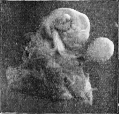
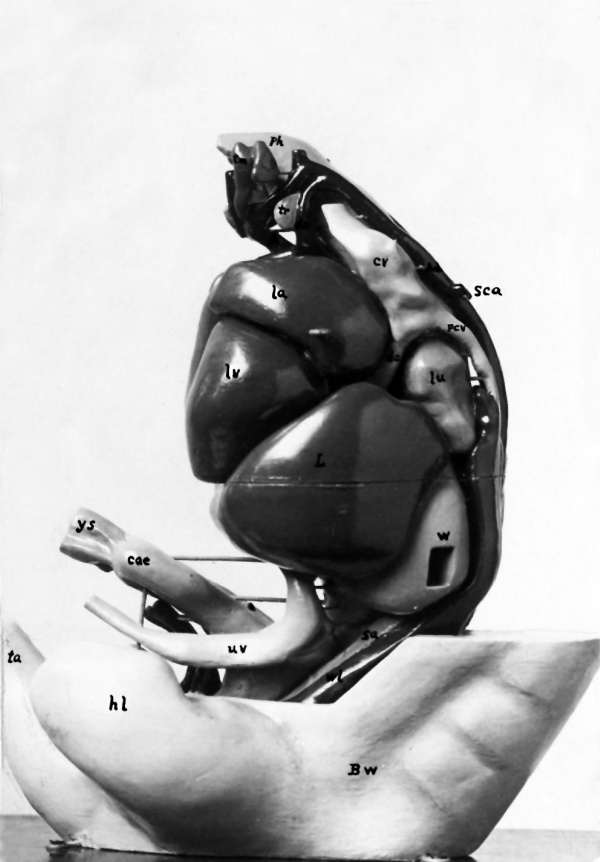
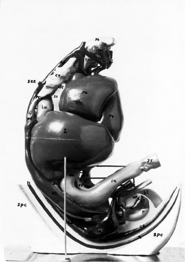
The embryo used for this study is catalogued as No. H60 in the collection of human embryos in the Anatomical Laboratory of the University of Missouri. It measured eleven millimeters in head-rump length, after fixation and hardening in alcohol, and was excellently preserved. According to MALIBS rule, it would be about 33 days old. Fig. 1 is from a photograph of the embryo, together with its yolk sac, membranes, etc. The embryo was stained in bulk in alum-cochiueal, embedded in paraffin, and cut in serial transverse sections 20,1; thick. In order to study the structure of this embryo, two models were reconstructed by B0nN’s wax plate method. The first, which is represented by Figs. 2 and 3, is a finished model carefully reconstructed by Mr. BONNOT to show the form and relations of the cervical, thoracic, abdominal and pelvic viscera, together with the large blood vessels. The second was a rough model reconstructed by Mr. SEEVERS in order to determine the volume of a) the entire embryo and b) the principal parts and organs of the embryo. The volumes were measured by the water—displacement method, and then the actual volume was computed by dividing the result by the cube of the enlargement (75 diameters).
Fig. 1. From a photograph of the 11 mm human embryo (No. H60), somewhat magnified, showing the right side of the embryo, with the umbilical cord, yolk sac, and membranes. The tip of the tail is turned somewliat to the left, and is hidden by the umbilical cord.
The following table shows the volume of the model and also the (computed) actual volume of the embryo and the various parts.
Table 1. Volume of Embryo N0. H60 (11 mm)
, Volume of Actual Part“ of Body I Model i Volume Entire embryo 41200 cc. 0,0976 cc. Head of embryo 182100 ,, 0,0-13-1 ,, Trunk (without limbs) 20 700 ,, 0,0491 ,, Upper extremities 1200 ,, 0,0028 ,, Lower extremities 1000 ,, 0,0023 ,, Brain 8350 ,, 0,0198 ,, Spinal cord 2000 ,, l 0,0047 ,, Lungs 150 ,, 0,0003E) ,, Heart 1 500 ,, ; 0,0035 ,, Liver 2000 ,, 0,0047 ,,
In the next table, the absolute volume and the percentage of the total volume of the body is given for the various parts of the embryo.
Table II. Volumes of Embryo, Newborn, and Adult
11 mm. Embryo Newborn Adult volume °/,, of volume "/0 of volume * °/0 of cc. whole ee. Whole cc. 1 whole Whole body 0,0976 100 1 3440_ 100 _ 00 000 100 Heart 0,0035 3,04 1 Eff I { 300 0,5 Lungs 000035 0,30 } ' 1542 2,57 Liver 0,0047 4,85 E r 1463 -2,44 Brain ; 0,0198 20,20 371,7 10,8 1314 2,19 Spinal cord , 0,0047 4,85) 3,9 0,11 25,1 0,04 Head 0,0434 44,42 Trunk 0,0-L91 50,12 Upper extremities ‘ 0,0028 2,91
Lower extremities V 0,0023 2,43 454
For purpose of comparison, some figures (quoted from various sources in V11«n:ou1)'r’s “l)aton und 'l‘al)ellen”, 2. Aufl., 1893, and in DONALDSON’s "Growth of the Brain”, 1895) are also given for the newborn and for the adult. These figures are not in all cases directly comparable with each other, but will serve to indicate approximately the relations. For the heart, lungs and liver of the newborn, two sets of measurements are quoted. The lungs in the first case were evidently in the unexpanded fetal condition.
Fig. 2. From a photograph of the left side of the model. showing the cervical, thoracic, and abdominal viscera, as well as the large blood vessels. In the lower part. of the model, a portion of the external body wall. including the lower extremity and tail, is shown. The original model is forty-seven centimeters high, being reconstructed with an enlargement of seventy—five diameters. For convenience, the model isgmade in two segments, the plane of division passing horizontally through the liver.
Explanation: A ascending aorta. a dorsal aorta. a‘ left aortic arch. ac anterior cardinal vein (sinus-like dilatation). c caecum, not marked externally, though its cavity is distinct internally. co colon. d ductus Cuvier. hI hind limb. l lung. L liver. la left auricle. lv left ventricle. m mesentery. pa posterior cardinal vein. ph pharynx. s somite (external surface). so anlagc of sexual gland. so origin of suhelavian artery. sr suprarenal body (slightly visible). ta tail. th thymus, including the main gland and also the smaller “nodulus thymicus”. tl lateral anlage of the thyroid gland, located between the fourth and fifth branehial arterial arches. tm, median thyroid anlage. u umbilical vein. w Wolffian body. x quadrangular window cut through the thick-walled great omentum into the bursa omentalis. The anlage of the imperfectly differentiated spleen lies in the oiuental wall just behind this window. Internal to the omentum lies the stomach (visible through the window in the original model, though not in the photograph). ys yolk stalk, out near attachment to intestinal loop.
DONALDSON also gives (upon the authority of F. P. MALL) the combined volume of brain and spinal cord in the human embryo of 2 weeks at 0,04 cc., of 4 weeks at 0,2 cc., andof 12 weeks at 3,0 cc.
Fig. 3. From a photograph of the right View of the model. On this side, the lower part of the body wall is not seen as on the left side, but has been dissected out to the mid-sagittal plane, giving a side view of the spinal cord, notochord, the descending aorta with its branches, and the pelvic viscera.
Explanation (in addition to letters shown in Fig. 2): a‘ right aortic arch. al allantois. b Wolffian duet. ca caudal artery. cl cloaca. d duodenum. ha hypogastrie artery. i intestine. n notochord. o gall bladder. r rectum. ra right auricle. rv right ventricle. sc spinal cord. sv sinus venosus. u ureter. Its T-shaped upper extremity, forming the anlage of the permanent kidney, is almost entirely hidden by the hypogastric artery. up urinogenital papilla. vv vitelline (omphalomesenteric) vein.
These figures for the embryos of 2 and 4 weeks are evidently too large, however, being apparently larger than the volumes of the entire embryos of those ages. ' ‘
It will be observed from the preceding table that the heart is relatively large in the 11 mm embryo, being relatively about 6 times as large (in volume) as in the newborn, and more than 7 times as large as in the adult.
The lungs, on the contrary, are relatively only 5’/7 as large in the embryo as the newborn lungs before expansion (volume 43,5 cc., or 1,26 0/0 of whole body), or 1/, as large as the newborn lungs after expansion (volume 90 cc., or 2,62 0/0). 'l‘l1e adult lungs, according to the figures given, appear to be of about the same relative size as the expanded lungs of the newborn.
The liver in this embryo appears to be relatively only slightly larger than in the two newborn specimens for which figures are given, but it is relatively twice as large as in the adult.
The brain in this embryo is seen to be relatively large. It is, however, not so large when compared with the newborn (about twice as large) as it is when compared with the adult (about 9 times as large).
The spinal cord, however, shows the most surprising figures. That it is relatively enormous in size is evident from the portion visible in the model (Fig. 3 sp). Its volume, i11 this embryo, is exactly the same as that of the liver, or 4,85 0/0 of the whole body. This is about 44 times as large, relatively, as in the newborn, and 115 times as large as in the adult! It is evident, moreover, when the figures for the brain and spinal cord are compared, that the brain falls off most rapidly in size (when compared with the entire body) between birth and adult life; whereas the spinal cord decreases most rapidly in relative size before birth. Comparing the newborn with the embryo, the whole body has increased about 35000 times in volume, the brain about 19000 times, and the spinal cord about 830 times. From the newborn to the adult, the whole body increases about 17,4 times, the brain nearly 3,5 times and the spinal cord 6,4 times in volume.
The head of the embryo is, as is well known, relatively very large, the trunk and extremities small. No exact figures for comparison with later stages are available, however.
In form and relations, the various organs in this embryo agree, in general, with those of corresponding embryos already described in the literature. The more important relations are shown in the two views (Figs. 2 and 3) of Mr. Bonnor’s model. A brief description of the various structures is given under the explanation of these figures.
The levels of the principal organs with respect to the vertebral column were observed, and are indicated in the following table. The levels at this stage are most readily determined by means of the corresponding spinal nerve roots. All the organs noted he at levels considerably higher than in later stages. 457
Table III. Vertebral Levels of Organs in Embryo H60 (11 mm)
| Organs | Vertebral Level |
|---|---|
| Heart | 6th cervical to 4th thoracic |
| Formation of dorsal aorta | between 7th cervical and 1st thoracic |
| Origin of subclavian art. | 7th cervical |
| Ducts of Cuvier | 7th cervical |
| Origin of vitelline art. | 8th thoracic |
| Oesophagus | 5th cervical to 3rd thoracic |
| Stomach | 3rd thoracic to 7th thoracic |
| Liver | 3rd thoracic to 7th thoracic |
| Trachea | 5th cervical to 1st thoracic |
| Lungs | 1st thoracic to 3rd thoracic |
| Thyroid gland | 5th cervical |
| Suprarenals | 3rd thoracic to 6th thoracic |
| Wolffian bodies | 2nd thoracic to 1st lumbar. |
Certain features concerning the blood vessels in this embryo deserve special mention. The dorsal aorta is formed by the union of the two lateral aortic arches, just below the origin of the subclavian (vertebral) arteries. At its origin, the aorta is comparatively narrow in caliber (Figs. 2 and 3), but gradually enlarges as it passes downward, until its diameter becomes at least twice as great towards its lower e11d (Fig. 3). The hypogastric and caudal arteries are seen to be wide at their origin, but soon diminish rapidly in caliber (Fig. 3).
The vitelline artery in this embryo arises from the aorta opposite the 8th thoracic vertebra, and runs downward in the mesentery of the U-shaped loop of the intestine. On reaching the extremity of the loop, the artery divides into two branches which encircle the intestine, uniting again into a single trunk at the attachment of the yolk stalk (Figs. 2 and 3ys). HOCHSTETTER (7) has described a similar ring formed by the vitelline artery in mammals (out).
In earlier embryonic stages, as is well known, there exist two distinct vitelline arteries passing out, one on each side of the intestine, to reach the yolk sac. One of these (usually the left) is said to atrophy, leaving the other as a single vessel. The arterial ring existing in this case does not agree with this theory, however. Possibly the single trunk is formed by a fusion of the two primitive vitelline arteries except where they persist to form this intestinal ring. Later, one side of the ring evidently atrophies, leaving a single vitelline artery. Still later, all of the vitelline artery distal to the intestine atrophies with the yolk sac, the proximal portion persisting as the superior mesenteric artery of the adult.
The vitelline (omphalomesenteric) vein crosses above the intestine at the attachment of the yolk stalk, and enters the mesentery, forming a prominent ridge on its upper (primitive left) surface. On approaching the duodenum, this ridge becomes still more prominent (Fig. 3 '01)), and at one place the peritoneum entirely surrounds the vessel, which at this point is free from the surface of the mesentery. The vein finally passes under the duodenum to enter the liver.
Just below the duodenum, a branch from the vitelline vein extends out into the mesentery, and in the sections is found to accompany the vitelline artery. This branch evidently represents the superior mesenteric vein of the adult. The vitelline vein (distal to the duodenum) already shows sign of involution. Its walls are thickened, and its lumen very narrow, in places almost obliterated. No such changes are seen in the vitelline artery, however.
That the vitelline (omphalomesenteric) vein does not persist as the superior mesenteric vein was noted long ago by LUSCHKA (10). He states that the omphalomesenteric vein 1) disappears in man in embryos of the third month, but can still be injected through the heart at birth in the carnivora which are born blind (dog, cat, etc.). ALLEN (1) confirmed these results in the dog, cat, lion, and guinea pig.
These results, however, have evidently been overlooked by many later writers. HIS (6) makes no mention of them in his elaborate work. He states that the omphalomesenteric vein becomes the superior mesenteric and the portal veins. The omphalomesenteric does become, in part, the portal vein, but evidently only a, small portion of it becomes superior mesenteric vein (i. e., the portion from the point where it is joined by the branch which does represent the true superior mesenteric up to the point where it receives the splenic vein, forming the portal vein).
MINOT (13) does not make a clear statement as to the relation of the vitelline vein to the superior mesenteric vein, and does not mention the atrophy of the vitelline vein. The same may be said of the text-books of MARSHALL (ll), MC MURRICH (12), YOUNG and Ronuvson (14), HERTWIG (5), and others.
DEXTER (3) describes, apparently as an original observation, the atrophy of the omphalomesenteric vein in the fetal cat, and an independent formation of the superior mesenteric vein. LEWIS (9) later verified this condition for the pig, crediting DEXTER with the original discovery.
Fosren and BALFOUR (4), however, describe the superior mesenteric vein in mammals as eventually joining the vitelline to form the portal vein. CIIARPY (2) also states the relation correctly, and credits LUSCHKA with the discovery. HOCHSTETTER (8) cites and endorses the statements of ALLEN (1).
- LUSCHKA also included the artery in his statement, but this is evidently an error.
The large sinus-like dilatations of the lower portion of the jugular (anterior cardinal) veins are very evident in the model (Figs. 2 and mm).
Finally it may be worthy of note that in this embryo the umbilical vein in two places divides and reunites. (The vein shown in the model is the left umbilical vein, the rudimentary right umbilical vein not being represented.) Below the distal portion of the intestinal loop, while still lying in the wall of the umbilical cord, the umbilical vein divides into two equal branches, which immediately reunite (not seen in lateral views of model). In the body wall below the liver (Fig. 212) the umbilical vein again divides, this time into three branches, one large and two small, which soon reunite into a single trunk before entering the liver.
Additional Plates
From Bonnet's 1906 thesis, model photographs that did not appear in this paper.
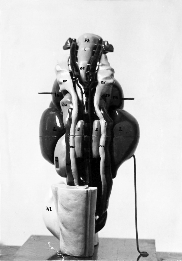
Plate 3. Photograph of the posterior view of the model
Showing the descending aorta, the cardinal veins, the thoracic and abdominal viscera. In this view the levels or the spinal nerve roots are indicated by short transverse stripes on the oesophagus and descending aorta. Ad, descending aorta; ov, anterior cardinal veins; hl, hind limb; la, left suriele; 3, liver; la, lune; oe, oesophagus; per, posterior oarlisal veins; ph, pharynx; ra, right auricle; s, suprarenals; sv, sinus venosus; window out in mesogastrium; wl Wolffian bodies.
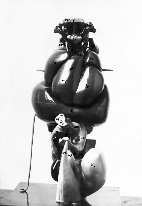
Plate 4. Photograph of the anterior view of the model
The small uper part shows the pharynx and its appendages, and the branchial arteries. The large middle part of the model represents the heart above and the liver below. Below the liver are the intestines, yolk stalk and umbilical vessels.
A, ascending aorta; cae, caocum; cv, anterior cardinal veins; H, heart; ha, hypogastric artery; hl, hind limb; ia. iliac artery; in, intestine; L, liver la, left auricle; lr, Larynx; lv, left ventricle; pa, pulmonary artery; ph, pharynx; rs, right auriole; rv, rimht ventricle; ea, sexual anlsge; t, tongue; ta, tail; tm, thyxus; tr, thyroid gland; up, urinogenital papilla; uv, umbilical vein; va, vitelline artery; vd, vétclline duet; vv, vitelline vein; wl, Wolffian bodies.
References to Literature
1) ALLEN, W., Omphalo-mesenterie Remains in Mammals. Journal of Anatomy and Physiology, Vol. 17, 1883. — Also reviewed in HOFMANN u. SeHwAL1m’s Jahresber., Bd. 11, 1883, p. 384-385.
2) CHARPY, A., in Poininii-CHARPY’s Traité d’anatomie humaine, T. 2, 3, Paris 1902, p. 1023.
3) DEXTER, F., The Vitelline Vein in the Cat. American Journal of Anatomy, Vol. 2, 1902, p. 261~—267.
4) FOSTER, M., and BALFOUR, F. M., The Elements of Embryology, London 1889.
5) HERWIG, 0., Textbook of Embryology. Translated by E. L. MARK, New—York 1892.
6) His, VV., Anatomie menschlicher Embryonen, Bd. 3, Leipzig 1885.
7) HOCHSTETTER, F., in HE1:TwIG’s Handbuch der Entwiekelungslehre der Wirbeltiere, Lief. 13—15, 1903, p. 141.
8) —, ibid., Lief. 3, 1902, p. 2]—166.
9) LEWIS, F. T., The gross Anatomy of a 12 mm Pig. American Journal of Anatomy, Vol. II, 1903, p. 211-225.
10) LUSCHKA, 11, Die Anatomie des Mensohen, Bd. II, Tiibingen 1863, p. 341.
11) MARSHALL, A. M., Textbook of Vertebrate Embryology, New York 1892.
12) McMurrich, J. P., The Development of the Human Body, Philadelphia 1902.
13) Minot, C. S., Human Embryology, New York 1892.
14) YOUNG, A. H., and ROBINSON, A., in Cunningham's Textbook of Anatomy, New York 1905.
Cite this page: Hill, M.A. (2024, April 28) Embryology Paper - On the structure of a human embryo eleven millimeters in length. Retrieved from https://embryology.med.unsw.edu.au/embryology/index.php/Paper_-_On_the_structure_of_a_human_embryo_eleven_millimeters_in_length
- © Dr Mark Hill 2024, UNSW Embryology ISBN: 978 0 7334 2609 4 - UNSW CRICOS Provider Code No. 00098G

