Paper - Further observations on the ossification of the human lower jaw
| Embryology - 18 Apr 2026 |
|---|
| Google Translate - select your language from the list shown below (this will open a new external page) |
|
العربية | català | 中文 | 中國傳統的 | français | Deutsche | עִברִית | हिंदी | bahasa Indonesia | italiano | 日本語 | 한국어 | မြန်မာ | Pilipino | Polskie | português | ਪੰਜਾਬੀ ਦੇ | Română | русский | Español | Swahili | Svensk | ไทย | Türkçe | اردو | ייִדיש | Tiếng Việt These external translations are automated and may not be accurate. (More? About Translations) |
Low A. Further observations on the ossification of the human lower jaw. (1909) J Anat Physiol. 44(1): 83–95. PMID 17232830
| Online Editor |
|---|
 Also by this author: Low A. Description of a human embryo of 13-14 mesodermic somites. (1908) J Anat Physiol. 42(3): 237-51. PMID 17232769 | PMC1289161
|
| Historic Disclaimer - information about historic embryology pages |
|---|
|
Further Observations on the Ossification of the Human Lower Jaw
By Alex. Low, M.A., M.B.
From the Anatomy Department, University of Aberdeen
With 1 Plate
Introduction

In, a research published in 1905, the appearances presented by serial sections of many stages of developing human lower jaw were described in detail by me. As regards human lower jaw I came to the following conclusions :—
- Each half of the lower jaw is developed in membrane as a single skeletal element—the dentarg/—and the so-called splenial element is simply an extension of this, helping to form the inner alveolar wall, and does not exist as a separate element.
- Meckel’s cartilage becomes ossified and incorporated with that part of lower jaw below and inside the mesial and lateral incisor teeth. Posterior to this point Meckel’s cartilage does not enter into the formation of lower jaw. The anterior extremities of Meckel’s cartilages also do not enter into lower jaw formation, but usually persist throughout foetal life as one or two cartilaginous nodules behind the symphysis.
- At a comparatively late stage in the development of the lower jaw at the third month of foetal life—certain accessory cartilaginous nuclei (“accessory cartilages”) appear in connection with the primary membrane bone. Thus there is a well-defined, wedge-shaped condylar cartilage and a smaller eoronmld cartilage. In addition to these there are also smaller cartilaginous nuclei along the margins of both alveolar walls in front as Well as along the front of the lower border of the jaw. In the human lower jaw I have not observed a definite angular nucleus, although in many other mammals a distinct and Well-defined angular cartilaginous nucleus exists. These various accessory cartilaginous nuclei do not indicate separate elements, but are an adaptation to the growth of the jaw.
In the present communication I do not enter into the historical aspect of lower jaw ossification, but simply state that of recent Workers on the subject Dieulafé and Herpin, Fawcett and Gaupp support these conclusions. On the other hand, K. von Bardeleben and H. Fuchs are still of opinion that the mammalian lower jaw is made up of more than one skeletal piece. K. Von Bardeleben argues that the chin in man and mammals is a special skeletal element —the os mentale— and that there are also found in the lower jaw condyloid, coronoid, angular, marginal, and dentale as separate elements. More recently H. Fuchs states that in the rabbit embryo there is a distinct and separate splenial element, that the condylar cartilage has its origin from Meckel’s cartilage, and further thinks my series of early human embryonic lower jaw not complete enough for settling such a point.
Henneberg gives a very excellent description of the appearances presented by the developing lower jaw in human foetuses from 40 mm. to 240 mm., crown—rump measurement. Dieulafé and Herpin have no stage between 18 mm. and 32 mm., and in Fawcett’s paper also there is given.no detailed description between the 21- and 42-mm. stage. In my -former paper, although I covered most of the stages, I also had no good stage between 19 and 28 mm. So that one admits that the stages roughly between 20 and 30 mm. are insufliciently described.
Since the publication of my previous paper I have examined serial sections of the heads of a large number of early human embryos. Through the great kindness of Professor Keibel of Freiburg I had the opportunity of examining serial sections of many embryos which he had in his possession in connection with the production of his Normentafel zur Entwicklungsgeschichte des Menschen. I have also added several series to my own collection. In this way it has been possible for me to study the ossification of the human lower jaw in a practically complete series of embryos from 15 to 30 mm. in length. From a study of these along with the older stages which I already have described in detail, we obtain a complete picture of the ossification of the human mandible.
Further, I have selected the more important stages of the developing mandible and made reconstruction wax-plate models by Born’s methodin all reconstructing five distinct stages. These models serve to demonstrate plainly the relation of Meckel’s cartilage and the “accessory cartilages” to the membrane bone of the jaw as well as the relative position of the various nerves.
Personal Observations
Meckel’s cartilage is present in a precartilaginous stage in an embryo of 12 mm., is cartilaginous in an embryo of 146 mm., and in an embryo of 15 mm. is well formed, passing -forwards towards the middle line, its ventral end almost meeting but not fusing with its fellow of the opposite side.
Ossification in the mandible is first observed in an 18-mm. embryo; that is, somewhat later than the appearance of ossification in the clavicle, which in Professor Keibel’s series is present at the 16—mm. stage—ossification being present in the clavicle in three embryos in which there is still no trace of ossification in the mandible. In an embryo 18 mm. in length ossification appears as a delicate lamella of bone developed in the mesenchyme on the outer aspect of the ventral extremity of Meckel’s cartilage. The bone extends backwards from near the middle line on the outer side of the cartilage and under the inferior dental nerve and its incisive branch.
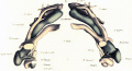
In another embryo of 18 mm. belonging to Professor Keibel’s series, in which ossification is further advanced, the relation of developing bone to cartilage is particularly well seen. Figs. 1 and 2 in the Plate represent views of a plate-model of the mandible of this embryo. Membrane bone is developed close on the outer aspect of the ventral end of Meckel’s cartilage and already forms a fairly stout single lamella extending from the symphysis backwards for about half the length of Meckel’s cartilage. In coronal sections, 15 microns thick, bone is present in ten sections before Meckel’s cartilage is reached, so that there is a considerable sheet of bone anterior to the cartilage. The bone lies rather below the level of the nerves, and seems to be deposited with regard to the inferior dental nerve and its branches. The upper margin of the bone already shows an oblique groove in which the mental nerve lies. The incisive nerve also lies on a slight groove on the upper margin of the bone, and a process of bone is growing up to the inner side, between the nerve and Meckel’s cartilage, forming an inner alveolar margin—the commencing so-called splemlal.

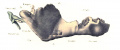
Fig. 1. Part of a coronal section of the head of a human embryo 18 mm. in length (Keibel’s Collection, No. 1421). x 25. The section passes through the region of the mouth and mandible, and shows the relations of Meckel's cartilage, the developing membrane bone of the lower jaw, and the mandibular nerve.
The course and shape of Meckel’s cartilage also seems to be largely modelled by the nerves which are relatively very large. Thus there is a bend inwards of the cartilage at the point where the posterior division of the inferior maxillary nerve comes into relation with it. This nerve—trunk breaks up on the upper border of the cartilage; the auriculo—temporal nerve passing backwards, closely applied to the lateral surface of the cartilage; the lingual and inferior dental nerves pass forward, the former closely applied to the inner aspect of the cartilage, the latter to the outer aspect. Where the lingual nerve passes in its final distribution to the tongue there is another sharp bend of the cartilage round the nerve. The ventral end of the cartilage not only bends inwards but upwards, at the same time becoming enlarged and flattened. The mylohyoid branch of the inferior dental nerve keeps close to the outer and then under aspect of the cartilage.

Fig. 2. Coronal sections of the right half of the lower jaw of a human embryo 24 mm. in length. x 30. I. shows the membrane bone in front of Meckel’s cartilage. II. is through the ventral extremity of Meckel’s cartilage. III. is a section through the mental foramen showing a bridge of bone behind the canine tooth-germ. IV. Through middle of the body of jaw. V. Through the coronoid process. J., membrane bone of lower jaw ; D., dental lamina; N., interior dental nerve; My., mylohyoid nerve ; L., lingual nerve ; M.C., Meckel’s cartilage; Cr., coronold process ; S., submaxillary gland.
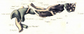
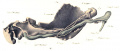
In an embryo 24 mm. in length the process of ossification has continued to extend so that the membrane bone now forms a sheet on the lateral aspect of Meckel’s cartilage from the symphysis in front to the auriculo-temporal nerve behind. figs. 3 and 4 in the Plate, copied from a plate-model of the mandible of this embryo, show the disposition of the parts. The bends in Meckel’s cartilage are more pronounced; there is a bend where the branches of the inferior maxillary nerve get in relation with it, and another more pronounced bend just inside the mental foramen, the cartilage here passing rapidly in towards the middle line and also rising up. The ventral end of the cartilage is now relatively larger and more expanded. The mental groove is now converted into a foramen, its margins having fused over the mental nerve. The angle and coronoid process are mapped out in membrane bone. The inner alveolar margin is- extending backwards so that coronal sections in front and behind the mental foramen are Y-shaped in appearance, there being thus a well-formed tooth gutter. The mylohyoid nerve passes close round the lateral aspect of the cartilage between it and the membrane bone.

Fig. 3. Coronal section of the lower jaw of a human embryo 28 mm. in length. x 25. The section passes through the region of the symphysis and shows the lamella of membrane bone lying to the outer aspect of the enlarged anterior extremity of Meckel’s cartilage.
In an embryo 28 mm. in length ossification has still further extended, but the relations and appearances are much the same as in the mandible of the last embryo. Each half of the lower jaw is now mapped out as one complete membrane bone with a dental shelf beginning to overhang Meckel’s cartilage. The cartilage still shows pronounced bends, and terminates in front in an enlarged, somewhat knobbed extremity, with the membrane bone on its outer aspect (fig. 3). There is no trace of commencing ossification in the cartilage. A plate-model was reconstructed from the serial sections of the mandible of this embryo, but except in that the bone has extended somewhat, the model presents very similar appearance to that of the 24-mm. embryo.
In an embryo 31 mm. in length the only changes are that the anterior extremities of Meckel’s cartilages are more flattened from before back, and at a point between the lateral incisor and canine tooth—germs the cartilage cells are becoming enlarged preparatory to ossification. In an embryo 36 mm. in length, at a corresponding point in Meckel’s cartilage, ossification is taking place in the perichondriuin on the upper and lateral aspects of the cartilage, and here the cartilage cells are enlarged and vacuolated.

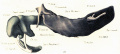
Fig. 4. Plate-model of the right half of the mandible of a. human foetus 43 mm. in length. The original model is thirty times enlarged, and A represents a view of the outer aspect, and B of the inner aspect reduced about one-third. Cy., condyle; Cr., coronoid process still formed of membrane bone; F.m., foramen mentale; M.C., Meckel's cartilage; X., point of commencing resorption in the cartilage; Sp., inner alveolar margin extending inwards from the primary membrane bone; Sy., symphysis; C.m., capitulum mallei ; M.m., manubrium mallei; Pr.F., processus Folianus (antr.).
In a foetus 43 mm. in length ossification is now taking place in Meckel’s cartilage where the cartilage is in close relation with the membrane bone.
Fig. 4 shows views from the outer and inner aspects respectively of a plate-model of the right half of the mandible of this foetus. From the outer aspect the various parts of the adult. jaw are readily recognised—the slit-like mental foramen, the well—marked coronoid process, the angle, the ramus and the condyle, the latter formed of condensed mesodermic cells with so far no trace of cartilage. On the inner aspect Meckel’s cartilage is seen running close inside the bone; behind, it is below the level of and quite distinct from the condyle, while anteriorly it is overhung by the inner alveolar shelf. The ventral end of the cartilage is expanded and flattened from before back.

Fig. 5. Coronal section of the left half of the lower jaw of a human foetus 65 mm. in length. x 50. The section passes through the canine tooth-germ, and shows Meckel’s cartilage partly surrounded by bone and undergoing resorption.
In a foetus 55 mm. in length the process of resorption and ossification of Meckel’s cartilage has extended backwards so far as the region of the canine tooth-germ. The cartilage cells are broken down and marrow cavities are being formed by osteoblastic tissue growing in from the closely applied membrane bone (fig. 5). The condyle, now distinctly outlined; shows commencing formation of cartilage—while the ascending ramus is still wholly formed of membrane bone. The coronoid process is thickened and formed of lattice-like membrane bone.
In a foetus 60 mm. in length the process of resorption and ossification of Meckel’s cartilage is more advanced, and the condyle is built up of hyaline cartilage. In a foetus 72 mm. in length Meckel’s cartilage is ossifying, and enclosed in a sheath of bone in a region corresponding to

Fig. 6. Coronal sections of the right half of the lower jaw of a human foetus 55 mm. in length. x30. I. Through the central incisor tooth-germ. II. Through the canine tooth germ. III. At mental foramen showing bridge of bone behind canine tooth-germ. IV. Through the first molar tooth-germ. V. Through the second molar tooth-germ. VI. Through the coronoid process. J ., lower jaw, :1 continuous piece of membrane bone; D., dental lamina and tooth-germs; N., inferior dental ' nerve; L.. lingual nerve: M.C., Meckel's cartilage; My., mylohyold muscle; S , submaxillary gland and its duct ; X., Meckel's cartilage undergoing resorption.
from the mental foramen to the front of the lateral incisor tooth—germ, while its anterior extremity remains unossified, passes close under the germ of the central incisor tooth, and meets its fellow of the opposite side. In a foetus 80 mm. in length the part of Meckel’s cartilage from the central incisor to the canine tootl1—germ is wholly taken into the membrane bone of the lower jaw. The condylar cartilage is well developed, and a strip of cartilage now appears along the anterior border of the coronoid process.

Fig. 7. Coronal section of the right half of the lower jaw of a human foetus 95 mm. in length. x 12. The section is through the coronoid rocess, and shows , the “accessory cartilage ” here. Near the lower border Meckel’s cartilage is seen lying apart ; at a corresponding point in the membrane bone is seen an interruption caused by the anteriorextremity of the condylar cartilage.
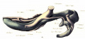
In a foetus 95 mm. in length the condylar and coronoid cartilages are both largely developed. figs. 5 and 6 in the Plate are copied from a platemodel of the right half of the mandible of this foetus. The positions and relations of the two main “accessory cartilages” of the mandible—the condylar and coronoid—are readily seen. The conolg/lwr cartilage is wedge shaped in appearance and passes downwards through the ascending ramus, its pointed lower extremity passing forward into the body of the jaw and terminating so far forward as the level of the front of the base of the coronoid process. The cartilage is covered on both lateral aspects by perichondrium continuous with the periosteum. Under the microscope the cartilage towards its anterior part shows regressive changes, being destroyed by giant cells, and its place taken by marrow tissue and eventually bone. Posteriorly the cartilage expands to form the condyle—formed of hyaline cartilage covered with perichondrium. The coronoid cartilage forms a well developed strip along the anterior border of the process. It also is covered with perichondrium continuous with the periosteum of the bone, and is being replaced by ossification extending into it from the neighbouring bone. In this specimen, in addition to these two main “accessory cartilages,” another accessory nodule is present on the inner aspect of the lower border of the mandible just outside the symphysis. Viewing the model on its inner aspect, Meckel’s cartilage is seen passing from behind forward, lying at first close below and inside the condyle, then descending it runs along below the inner alveolar shelf, gaining the lower border of the jaw just in front of mental foramen and under the canine tooth; at this point it passes into the jaw, and its further course is indicated by a bony ridge which ascends toward the symphysis under the central incisor tooth, the ventral end of Meckel’s cartilage again reappearing as a small piece of cartilage at the side of the symphysis.
In a foetus 103 mm. in length serial sections present practically the same appearances as in the last specimen, with the addition of a small cartilaginous nucleus along the inner alveolar margin in the region of the second incisor tooth. In (L foetus 130 mm. in length there is much increase in the size of the jaw as a whole. The condylar and coronoid cartilages are rapidly becoming ossified, and are relatively less in extent. In addition to these two “accessory cartilages ” there are small cartilaginous nuclei along both alveolar margins in the region of the incisor teeth as well as along the lower border of the jaw in front. In at faetus 210 mm. in length these different pieces of cartilage are still present, but are much smaller. In a faetns 230 mm. in length only traces of these cartilages remain, and they are now surrounded by bone. The cartilage in the coronoid process has practically disappeared, but the condylar cartilage still persists, although its anterior part is much broken up by large marrow spaces and deposits of bone. Meckel’s cartilage still exists behind the canine tooth.
From an examination of microscopic sections and naked-eye preparations of foetal lower jaw from this stage till full time one finds that the jaw rapidly becomes wholly converted into bone, Meckel’s cartilage behind the canine tooth gradually atrophies, all trace of the coronoid cartilage disappears, while remains of the condylar cartilage persist until birth.
These further observations support my previous conclusion that each half of the lower jaw is developed in membrane as a single skeletal piece— the dentamy. Meckel’s cartilage takes only a slight part in the formation of the mandible, and does not give rise to the condylar cartilage. There are certain “accessory cartilages ” in connection with the primary membrane bone, and the View may be taken that these represent separate skeletal elements. Against this view it is to be noted that these “accessory cartilages ” are late in appearing and, as Kolliker pointed out, are not in any way connected with the primordial cranium. Gaupp has also drawn attention to the fact that in connection with other membrane bones of the skull accessory cartilaginous nuclei of the same nature are developed. Further “accessory cartilages” have no separate centres of ossification. These cartilages are replaced by undergoing regressive changes, being invaded by the surrounding membrane bone, destroyed by giant cells, and replaced by marrow tissue and trabeculae of bone.
In conclusion, I wish to express my thanks to Professor Keibel of F reiburg University for afi"ording me every facility in connection with the construction of my wax-plate models and for the opportunity of studying his series of early human embryos. I also again express my indebtedness to Professor Reid for the opportunity of carrying on research work, and to the Carnegie Trustees for a grant toward the cost of the illustration of this paper.
Bibliography
vox BARDELEBEN, KARL, “ Der Unterkiefer der Siiugetiere, besonders des Menschen,” Anat. Anzeiger, Bd. xxvi., No. 4-5, p. 104, 1905.
BAUMULLER, B., “ Ueber die letzten Vertinderungen des Meckel’schen Knorpels,” Zeitsc/Lri'ft_/'1Zr wissen. Z0olog., Bd. xxxii., p. 466, 1879.
Bnocx, J ., “ Ueber die Entwickelung des Unterkiefers der Saugetiere,” Zeitschri/'t fair wissensclzaft. Zoologie, Bd. xxvii., p. 287, 1876.
BROMAN, IVAR, “Die Entwickelungsgeschichte der Gehorknochelclien beim Menschen”: lnaug. Diss., Lund, 1899 (Anat. Hejfte, Bd. xi., H. 4).
CALLENDER, G. D., “The Formation and Early Growth of the Bones of the Human Face,” Phil. ’['runs., vol. elix. p. 163, 1869.
l)xEUr.Am§: et HERPIN, “Développement de 1’os maxillaire inférieur,” Journal de l’Anatomie et de la Physiologic, vol. xlii. p. 239, 1906.
DIEULAFE et HERPIN, “Histogénése de l’0s maxillaire inférieur,” Journal de l’Anatomie et de la Physiologie, vol. xliii. p. 580, 1907.
FAWCETT, E., “The Ossification of the Lower Jaw in Man,” Proceedings of the Anat. Soc. of Great Britain and Ireland, p. 47, 1905.
FAWCETT, E., “Ossification of the Lower Jaw in Man,” The Journal of the American Medical Association, vol. xlv. p. 695, 1905.
FUCHS, H., “Bemerkungen iiber die Herkunft und Entwiekelung der Gehorkn'(5chelchen bei Kaninchen-Embryonen (nebts Bemerkungen iiber die Entwickelung des Knorpelskeletes der beiden ersten Viscera1bogen),” Archiv f. milcr. Anat., Supplement-Band, 1905.
GAUPP, E., “ Ontogenese und Phylogenese des schalleitenden Apparates bei den Wirbeltieren,” Er_r/ebnisse der Anatomie and Entwic/celungsgeschichte, vol. viii. p. 990, 1898.
GAUPP, E., “Die Nicht-Homologie des Unterkiefers in der Wirbeltierreihe,” Verhandlangen der anatomischen Gesellsc/zaff, 1905, p. 125. 94 Mr Alex. Low
GAUPP, E., “Died Entwickelung des Kopfskelletes,” Handbuch der vergleich. u. experiment. Entwickeiungslehre der Wirbeltiere, herausgegeben Von Dr 0. Hertwig, 1905.
HENNEBERG, 15., “Beitriige zur Entwickelungsgeschichte des Unterkiefers beim Menschen ”: Inaugural Dissertation, Berlin, 1894.
HUMPHRY, G., “ 011 the Growth of the Jaws,” Phil. Trans, vol. ix., 1871.
J ULIN, C., “ Recherches sur l’ossification du niaxillaire inférieur,” Arch. d. Biolog/., Tome i., p. 75, 1880.
KEIBEL, F. , Normentafel zur Entwickelungsgesr-hie}:te des Menschen.
Low, A., “The Development of the Lower Jaw in Man,” Proc. of the Anac. and Anthrop. Soc. of the University of Aberdeen, 1905.
Low, A., “The Development of the Lower Jaw in Man,” Proc. of the Anat. Soc. of Great Britain and Irelarzd, p. 26, 1905.
MAGITOT et ROBIN, “ Mémoirc sur un organe transitoire de la vie foetale désigné sous le nom de cartilage de Meckel,” Annales des sciences naturelles : Zoologie, Tome xviii., p. 213, 1862.
MASQUELIN, H., “Recherches sur le développement du maxillaire inférieur de 1’homme,” Bull. de l’Acad. reg. de Belgique, 2° série, Tome xlv., 1878. ,
MIES, J ., “Ueber die Knochelchen in der Symphyse des Unterkiefers vom neugebornen Menschen,” Anat. Anzeiger, 1893.
MECKEL, J. F., Handbuch der mehschlichen Anatomie, Bd. iv., Halle u. Berlin, p. 47, 1820.
PARKER, W. K., “On the Structure and Development of the Skull in the Pig,” Phil. Trans, vol. clxiv. p. 312, 1874.
QUAIN, Elements of Anatomy, London, vol. ii., part i., p. 78, 1899.
RAMBAUD et RENAULT, Origine et «léveloppement des as, Paris, 1864.
SCI-IAFFER, J ., “Die Verkniicherung des Unterkiefers und die Metaplasifrage, Archivfzir mi/croslwp. Anatomie, Bd. xxxii., p. 266, 1888.
STIEDA, L., “Studien iiber die Entwicklung der Knochen und des Knochen— gewebes,” Archiv f. mikr. Anat., Bd. xi., p. 235, 1875.
Srmsnzorr, Z. J., “Ueber die Histogenese der Knochen,” Untersuchungen aus dem pathol. Institutzu Zurich, I. Heft, Leipzig, p. 45, 1873.
BLAND SUTTON, J ., “Development of the Inferior Maxil1a,” Trans. of the Odontological Society, vol. xxv., New Series, p. 157, 1883.
TESTUT, L., Traité d’Anatom.ie Ilumaine, Quatrieme édition, Tome i., p. 216, 1899.
WOLFE, J., “Ueber das Wachstnm des Unterkiefers,” Arch. f. path. Anat. u. Phg/s., Bd. cxiv., p. 493, 1888.
Explanation of Plate

Figs. 1 and 2. Plate-model of mandible of human embryo 18 mm. in length (Keibel’s Collection, No. 1421). The original model is 50 times enlarged, and fig. 1 represents the model as seen from above and fig. 2 as seen from the right - both drawings reduced about one-fourth.
Figs. 3 and 4. Plate-model of mandible of human embryo 24 mm. in length. The original model is 50 times enlarged, and the drawings represent the right half of the model viewed from the outer and inner aspects, reduced about one-third.
Figs. 5 and 6. Plate-model of the right half of the mandible of a human foetus 95 mm. in length. The original model is 33 times enlarged and the drawings represent views of the outer and inner aspects reduced to one-third.
human embryo 18 mm mandible above
human embryo 18 mm embryo mandible right
human embryo 24 mm mandible right half outer aspect
human embryo 24 mm mandible right half inner aspect
human foetus 95 mm mandible right half outer aspect
human foetus 95 mm mandible right half inner aspect


Abbreviations
C. unpaired cartilaginous nodule behind symphysis.
Cu. accessory cartilaginous nodule.
Capit. mall. capitulum mallei.
Cart. Meckel. Meckel’s cartilage.
Cart. Reichert. Reichert’s cartilage.
Ch. ty. N. chorda tympani.
Cr. br. incud. crus brevis incudis.
Cr. long. incud. crus longum incudis.
Cr. coronoid accessory cartilage.
Cy. condylar accessory cartilage.
For. ment. foramen mentale.
Manub. mall. manubrium mallei.
N. auric. N. auriculo-temporalis.
N. facial. N. facialis.
N. mandib. N. mandibularis.
N. ling. N. lingualis.
N. ment. N. mentalis.
IV. mylo. N. mylo~hyoideus.
N. trigem. N. trigeminus.
Us. dent. Dentale.
Pros. Fol. processus Folianus (antr.).
Sy. symphysis.
| Historic Disclaimer - information about historic embryology pages |
|---|
|
Cite this page: Hill, M.A. (2026, April 18) Embryology Paper - Further observations on the ossification of the human lower jaw. Retrieved from https://embryology.med.unsw.edu.au/embryology/index.php/Paper_-_Further_observations_on_the_ossification_of_the_human_lower_jaw
- © Dr Mark Hill 2026, UNSW Embryology ISBN: 978 0 7334 2609 4 - UNSW CRICOS Provider Code No. 00098G