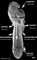
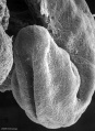
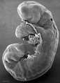
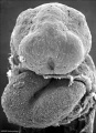
Human Embryo - Scanning electron microscopy
Introduction
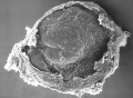
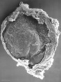
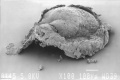
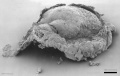
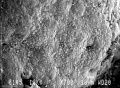
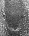
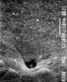
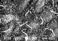
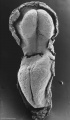
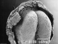
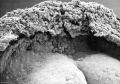
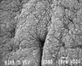
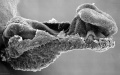
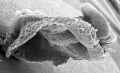
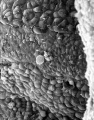
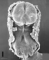
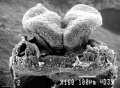
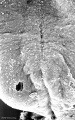
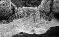
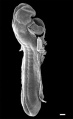
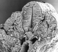
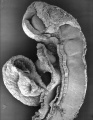
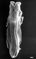
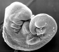
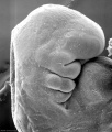
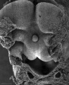
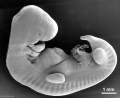
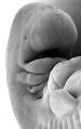
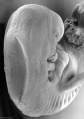
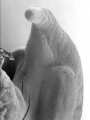
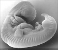
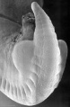
This page displays in gallery mode human embryos imaged using a scanning electron microscope. Currently only embryos up to stage 14 are available.
Image Source: Scanning electron micrographs of the Carnegie stages of the early human embryos are reproduced with the permission of Prof Kathy Sulik, from embryos collected by Dr. Vekemans and Tania Attié-Bitach. Images are for educational purposes only and cannot be reproduced electronically or in writing without permission.
- Links: Category:Scanning EM
Embryo Stages
Stage 7
- Stage7-sem6.jpg
- Stage7-sem7.jpg
- Stage7-sem8.jpg
- Stage7-sem9.jpg
- Stage7-sem10.jpg
- Stage7-sem11.jpg
- Stage7-sem12.jpg


Stage 8
- Stage8 sem8.jpg
- Stage8 sem9.jpg
- Stage8 sem10.jpg
- Stage8 sem11.jpg
- Stage8 sem12.jpg
- Stage8 sem13.jpg
- Stage8 sem14.jpg
- Stage8 sem15.jpg


Stage 9
- Stage9 sem8.jpg
- Stage9 sem9.jpg
- Stage9 sem10.jpg
- Stage9 sem11.jpg
- Stage9 sem12.jpg


Stage 10


Stage 11
- Stage11 sem1.jpg


- Stage11 sem12.jpg
- Stage11 sem14.jpg
- Stage11 sem15.jpg


Stage 12


Stage 13
- Stage13 sem4.jpg
- Stage13 sem5.jpg
- Stage13 sem6.jpg
- Stage13 sem7.jpg
- Stage13 sem8.jpg
- Stage13 sem9.jpg
- Stage13 sem10.jpg


Stage 14
- Stage14 sem7.jpg
- Stage14 sem8.jpg
- Stage14 sem9.jpg
- Stage14 sem10.jpg
- Stage14 sem11.jpg
- Stage14 sem12.jpg


External Links
External Links Notice - The dynamic nature of the internet may mean that some of these listed links may no longer function. If the link no longer works search the web with the link text or name. Links to any external commercial sites are provided for information purposes only and should never be considered an endorsement. UNSW Embryology is provided as an educational resource with no clinical information or commercial affiliation.
Glossary Links
- Glossary: A | B | C | D | E | F | G | H | I | J | K | L | M | N | O | P | Q | R | S | T | U | V | W | X | Y | Z | Numbers | Symbols | Term Link
Cite this page: Hill, M.A. (2026, July 4) Embryology Human Embryo - Scanning electron microscopy. Retrieved from https://embryology.med.unsw.edu.au/embryology/index.php/Human_Embryo_-_Scanning_electron_microscopy
- © Dr Mark Hill 2026, UNSW Embryology ISBN: 978 0 7334 2609 4 - UNSW CRICOS Provider Code No. 00098G