File:Wheeler024.jpg

Original file (592 × 700 pixels, file size: 49 KB, MIME type: image/jpeg)
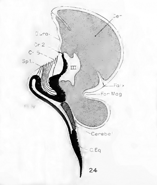
Fig. 24. Diagram of those structures of the central nervous system
Which lie near the midline and which can be identified. The outline of the subdural space used was obtained from the sagittal section.
Posteriorly this passes near to the median margin of the left encephalocele.
The cerebrum designated by a dotted line is shown protruding below the foramen magnum into the encephalocele.
A small portion of the cerebellum, represented by line-hatching, is seen to lie very much flattened on top of the cord.
The brain-stem and cord, much bent, arc shown in solid black.
Those cranial nerves which were identified are shown by lines.
Only the first spinal nerve is shown.
The floor of the fourth ventricle lies inverted on top of a flat cord.
File history
Click on a date/time to view the file as it appeared at that time.
| Date/Time | Thumbnail | Dimensions | User | Comment | |
|---|---|---|---|---|---|
| current | 09:11, 16 February 2011 | | 592 × 700 (49 KB) | S8600021 (talk | contribs) | ==Fig. 24. Diagram of those structures of the central nervous system== Which lie near the midline and which can be identified. The outline of the subdural space used was obtained from the sagittal section. Posteriorly this passes near to the median mar |
You cannot overwrite this file.
File usage
The following page uses this file: