File:Primordial germ cell DNA methylation 01.jpg

Original file (687 × 1,000 pixels, file size: 70 KB, MIME type: image/jpeg)
Primordial Germ Cell DNA Methylation
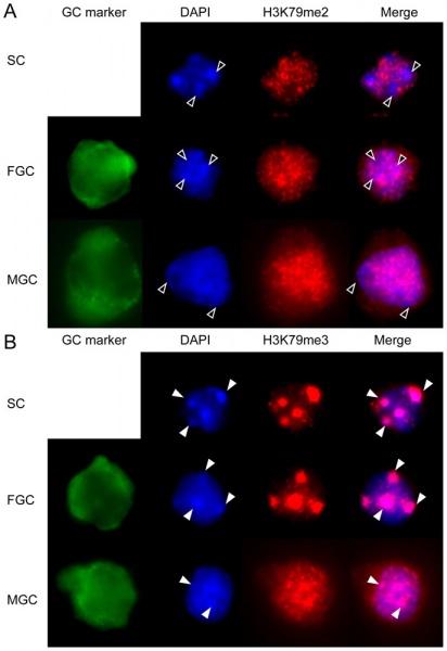
Nuclear distribution of H3K79me2 and H3K79me3 in fetal germ cells.
- H3K79me2 and H3K79me3 are associated with the DNA-unmethylated and methylated alleles of differentially methylated regions (DMRs)
Female and male germ cells (FGC and MGC, respectively) and female somatic cells (SC) at 15.5 dpc were stained with the H3K79me2 and H3K79me3 antibodies as indicated and are shown in the different channels separately and in red-blue composite.
(A) DAPI-stained chromocenters are excluded from H3K79me2 signals (open arrowheads).
(B) DAPI-rich chromocenters colocalize with H3K79me3 signals (closed arrowheads).
Figure 9. Journal.pone.0023848.g009.jpg
Reference
Abe M, Tsai SY, Jin SG, Pfeifer GP & Szabó PE. (2011). Sex-specific dynamics of global chromatin changes in fetal mouse germ cells. PLoS ONE , 6, e23848. PMID: 21886830 DOI.
Copyright
© 2011 Abe et al. This is an open-access article distributed under the terms of the Creative Commons Attribution License, which permits unrestricted use, distribution, and reproduction in any medium, provided the original author and source are credited.
Cite this page: Hill, M.A. (2024, April 26) Embryology Primordial germ cell DNA methylation 01.jpg. Retrieved from https://embryology.med.unsw.edu.au/embryology/index.php/File:Primordial_germ_cell_DNA_methylation_01.jpg
- © Dr Mark Hill 2024, UNSW Embryology ISBN: 978 0 7334 2609 4 - UNSW CRICOS Provider Code No. 00098G
File history
Click on a date/time to view the file as it appeared at that time.
| Date/Time | Thumbnail | Dimensions | User | Comment | |
|---|---|---|---|---|---|
| current | 00:30, 23 September 2011 |  | 687 × 1,000 (70 KB) | S8600021 (talk | contribs) | ==Primordial Germ Cell DNA Methylation== Nuclear distribution of H3K79me2 and H3K79me3 in fetal germ cells. Female and male germ cells (FGC and MGC, respectively) and female somatic cells (SC) at 15.5 dpc were stained with the H3K79me2 and H3K79me3 anti |
You cannot overwrite this file.
File usage
The following 2 pages use this file: