File:Pig - uterine epithelium SEM.jpg
From Embryology
Pig_-_uterine_epithelium_SEM.jpg (800 × 598 pixels, file size: 118 KB, MIME type: image/jpeg)
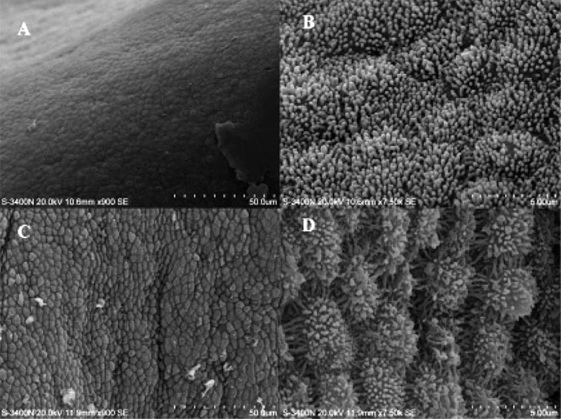
Pig - uterine epithelium SEM
Scanning electron microscope images of the endometrial surface of a Day 13 pregnant sow. (A) and (B) Tissue from between attachment sites. (C) and (D) Tissue at attachment sites.
- uterodomes (pinopods) Cellular features seen on the apical uterine epithelium surface, these micro-protrusions inter-digitate with microvilli on the apical syncytiotrophoblast surface of the blastocyst during adplantation and implantation process.
- Links: Image Pig SEM 1 | Pig Development | Image Human low SEM 1 | Image Human high SEM 2 | Implantation | Uterus Development
Reference
<pubmed>20640155</pubmed>| PMC2904919
Copyright
This article is an open-access article distributed under the terms and conditions of the Creative Commons Attribution license (http://creativecommons.org/licenses/by/3.0/).
Original File Name: Figure 1. 1471-213X-10-88-3.jpg
File history
Click on a date/time to view the file as it appeared at that time.
| Date/Time | Thumbnail | Dimensions | User | Comment | |
|---|---|---|---|---|---|
| current | 14:19, 19 October 2010 |  | 800 × 598 (118 KB) | S8600021 (talk | contribs) | ==Pig - uterine epithelium SEM== Scanning electron microscope images of the endometrial surface of a Day 13 pregnant sow. (A) and (B) Tissue from between attachment sites. (C) and (D) Tissue at attachment sites. :Links: Pig Development Original Fi |
You cannot overwrite this file.
File usage
The following 2 pages use this file:
