File:Nanagas1925-fig01d.jpg
Original file (600 × 765 pixels, file size: 45 KB, MIME type: image/jpeg)
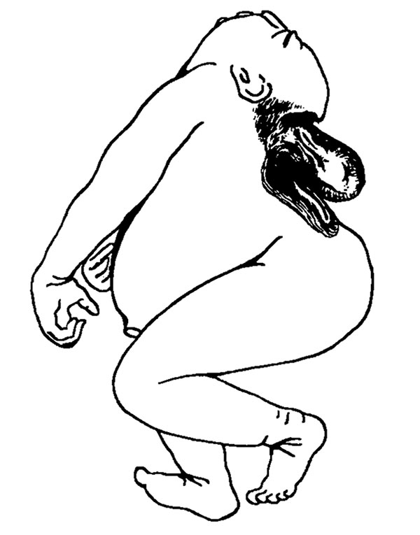
Fig. 1d. Microcephalic Craniorhachischisis
In this condition the spinal arches are separated by a wide cleft which usually extends to tlie lumbar region. The imperfectly developed brain in most of the cases is dislocated downward in the cervical region (hernia cerebri cervicalis) arid in a few instances this hernial formation is found in the thoracic region. The covering of the hernial sac is a meningeal-like membrane continuous with the covering of the flattened internal base of the cranium and that of the open vertebral arches. This type forms the largest number in our series of acrania, there being nineteen cases, or 83.3 per cent of the whole number of anencephalic cases.
| Historic Disclaimer - information about historic embryology pages |
|---|
 |
- Links: Fig 1. Anencephalus types | Fig 1a. anencephalic acranius | Fig 1b. anencephalic craniorhachischisis | Fig 1c. microcephalic acrauius | Fig 1d. microcephalic craniorhachischisis | Fig 1e. exocephalic acranius | Fig 16. anencephalic and normal fetuses | Historic Embryology Papers | Neural Abnormalities | Folic Acid and Neural Tube Defects | Skull Development
Reference
Nañagas JC. A comparison of the growth of the body dimensions of anencephalic human fetuses with normal fetal growth as determined by graphic analysis and empirical formulae. (1925) American J. Anatomy. 455-494.
Cite this page: Hill, M.A. (2024, April 26) Embryology Nanagas1925-fig01d.jpg. Retrieved from https://embryology.med.unsw.edu.au/embryology/index.php/File:Nanagas1925-fig01d.jpg
- © Dr Mark Hill 2024, UNSW Embryology ISBN: 978 0 7334 2609 4 - UNSW CRICOS Provider Code No. 00098G
File history
Click on a date/time to view the file as it appeared at that time.
| Date/Time | Thumbnail | Dimensions | User | Comment | |
|---|---|---|---|---|---|
| current | 12:23, 16 September 2015 | | 600 × 765 (45 KB) | Z8600021 (talk | contribs) | {{Nanagas1925 figures}} |
You cannot overwrite this file.