File:Mouse posterior neuropore Axd mutant.jpg

Original file (475 × 1,074 pixels, file size: 95 KB, MIME type: image/jpeg)
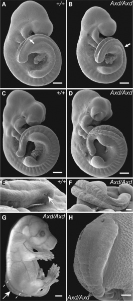
Failure of Posterior Neuropore closure in Axd Mouse Mutant Embryos
(A, C and E) Scanning electron microscopy of wild-type embryos reveals that posterior neuropore closure has progressed beyond the last formed somite and is at the level of the hindlimb bud at the 24 somite stage (arrow in A); by the 31 somite stage (C and E), the PNP has reduced in size to a small slit (arrow in E).
(B, D and F) In contrast, Axd mutant embryos exhibit dramatically enlarged PNPs at both 26 and 31 somite stages (B and D, respectively), in which closure does not progress beyond the level of somite 24 (arrow in B). At the 26 somite stage (B), the neural folds of the mutant embryo appear elevated, although closure has not progressed. By the 31 somite stage, the neural folds are splayed wide apart (D and F).
(G and H) In an Axd mutant fetus at E16.5, open spina bifida is evident (arrow in G) affecting the low thoracic, lumbar and sacral regions.
Transverse sections at the levels indicated by dashed lines in (G) are included in Supplementary Material, Fig. S1.
Scale bars: 500 μm (A–D), 100 μm (E), 200 μm (F), 2 mm (G) and 1 mm (H).
Reference
<pubmed>21262862</pubmed>| PMC3063985
Copyright
This is an Open Access article distributed under the terms of the Creative Commons Attribution Non-Commercial License (http://creativecommons.org/licenses/by-nc/2.5), which permits unrestricted non-commercial use, distribution, and reproduction in any medium, provided the original work is properly cited.
Original file name: Figure 1. Ddr03101.jpg
File history
Click on a date/time to view the file as it appeared at that time.
| Date/Time | Thumbnail | Dimensions | User | Comment | |
|---|---|---|---|---|---|
| current | 23:44, 2 May 2011 |  | 475 × 1,074 (95 KB) | S8600021 (talk | contribs) | ==Failure of Posterior Neuropore closure in Axd Mouse Mutant Embryos== (A, C and E) Scanning electron microscopy of wild-type embryos reveals that PNP closure has progressed beyond the last formed somite and is at the level of the hindlimb bud at the 24 |
You cannot overwrite this file.
File usage
The following page uses this file: