File:Mouse REN expression 01.jpg

Original file (904 × 1,000 pixels, file size: 293 KB, MIME type: image/jpeg)
Mouse Embryo REN Expression
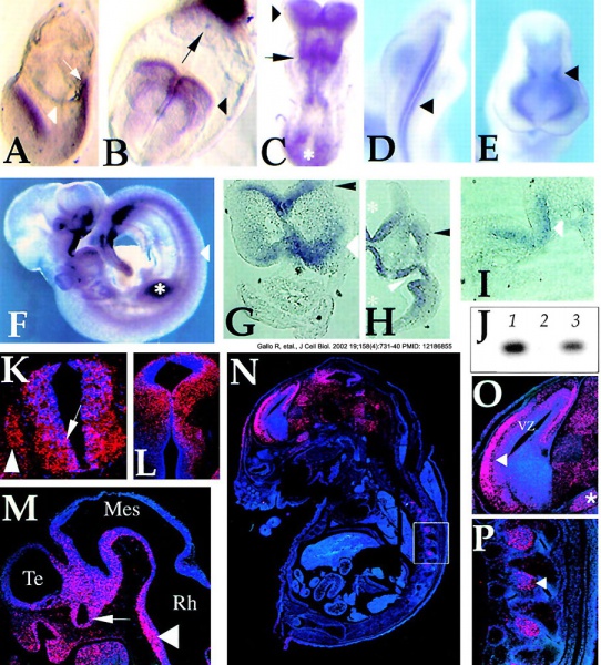
Whole-mount in situ hybridization at E7.5 (A), E8 (B), E8.25 (C), E9 (D and E) and E9.5 (F) reveals expression in the developing neural folds (A, white arrowhead in; B and C, black arrowhead), along the neural tube (D and E, black arrowhead), in the primitive streak (A, arrow; C, asterisk), in the ectoplacental cone (B, arrow), in newly forming somites and in the first rhombomere (C, arrow). At later stage (F), expression is restricted to the ventro-medial region of the neural tube (arrowhead), somites, optic and otic vesicles, the regions of the first branchial arch, the olfactory placode and developing limb buds (asterisk). Bright-field views of sections from whole mount (G–I) and postembedded in situ radioactive hybridizations (K–P) are also shown. Transverse sections confirmed expression in the cephalic neural folds (prospective forebrain; G, black arrowhead; E8.25), in the neuroepithelium of prospective hind brain (G, white arrowhead) and more caudally in the ventro-medial region of the neural tube (I, arrowhead; stage E8.25; K, arrow; stage E10.5). Coronal (H, stage E9.5) and sagittal (M, stage E10.5) sections show REN expression in the neuroepithelium of the fourth ventricle and of the diencephalon (H, asterisks) of the rhomboencephalic (Rh), mesencephalic (Mes), and telencephalic (Te) vesicles (M, arrowhead), in the olfactory placode including the epithelium lining the olfactory pit (M) and in the optic vesicle and stalk (H, white arrowhead; M, arrow). At later stages, REN expression characterizes more defined territories within the outer neuropeithelium layers of the midbrain and of the ventricular zone (VZ) of the cortical plate (L at stage E12.5; N; O, arrowhead; at stage E16.5). Some low and diffuse REN staining is also observed in the whole brain area (N). In the E16.5 embryo, outside of the brain REN expression was detected in dorsal root ganglia (N; P, arrowhead), preceded by expression in neural crest-derived spinal primordia (K, arrowhead). REN is also expressed in the trigeminal ganglion (O, asterisk) and in mesenchime of the maxillary component of the first branchial arch containing trigeminal neural crest tissue (H, black arrowhead). (J) RT-PCR analysis of REN expression in primary cultures of neural cells explanted from E9.5 embryonal neural tubes, cultured for 24 h (lane 1) and in differentiated N2a cells (lane 3), whereas no expression is detected in thymocytes isolated from newborn mice (lane 2).
Reference
<pubmed>12186855</pubmed>| PMC2174014 | J Cell Biol.
Copyright
Rockefeller University Press - Copyright Policy This article is distributed under the terms of an Attribution–Noncommercial–Share Alike–No Mirror Sites license for the first six months after the publication date (see http://www.jcb.org/misc/terms.shtml). After six months it is available under a Creative Commons License (Attribution–Noncommercial–Share Alike 4.0 Unported license, as described at https://creativecommons.org/licenses/by-nc-sa/4.0/ ). (More? Help:Copyright Tutorial)
File history
Click on a date/time to view the file as it appeared at that time.
| Date/Time | Thumbnail | Dimensions | User | Comment | |
|---|---|---|---|---|---|
| current | 13:32, 3 May 2013 |  | 904 × 1,000 (293 KB) | Z8600021 (talk | contribs) | ==Mouse Embryo REN expression== Whole-mount in situ hybridization at E7.5 (A), E8 (B), E8.25 (C), E9 (D and E) and E9.5 (F) reveals expression in the developing neural folds (A, white arrowhead in; B and C, black arrowhead), along the neural tube (D a... |
You cannot overwrite this file.
File usage
There are no pages that use this file.