File:Kollmann643.jpg
Kollmann643.jpg (556 × 491 pixels, file size: 48 KB, MIME type: image/jpeg)
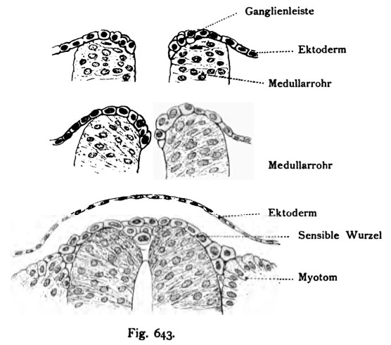
Fig. 643. Layout of the dorsal root ganglia of Human embryo of 13 somites, 2.5 mm long, 14 - 16 days
(After Lenhoss of ^ k.)
The ectodermal cells strip bar called ganglia merges with the other side, then the production of the sensory ganglion to- runways. The cell strips constricted off from the ectoderm and develops to the spinal ganglia, which in the gap between neural tube and Provertebral sinking. Spinal ganglia on further development stage see in the figures 642, 646, 647
- This text is a Google translate computer generated translation and may contain many errors.
Images from - Atlas of the Development of Man (Volume 2)
(Handatlas der entwicklungsgeschichte des menschen)
- Kollmann Atlas 2: Gastrointestinal | Respiratory | Urogenital | Cardiovascular | Neural | Integumentary | Smell | Vision | Hearing | Kollmann Atlas 1 | Kollmann Atlas 2 | Julius Kollmann
- Links: Julius Kollman | Atlas Vol.1 | Atlas Vol.2 | Embryology History
| Historic Disclaimer - information about historic embryology pages |
|---|
| Pages where the terms "Historic" (textbooks, papers, people, recommendations) appear on this site, and sections within pages where this disclaimer appears, indicate that the content and scientific understanding are specific to the time of publication. This means that while some scientific descriptions are still accurate, the terminology and interpretation of the developmental mechanisms reflect the understanding at the time of original publication and those of the preceding periods, these terms, interpretations and recommendations may not reflect our current scientific understanding. (More? Embryology History | Historic Embryology Papers) |
Reference
Kollmann JKE. Atlas of the Development of Man (Handatlas der entwicklungsgeschichte des menschen). (1907) Vol.1 and Vol. 2. Jena, Gustav Fischer. (1898).
Cite this page: Hill, M.A. (2024, April 26) Embryology Kollmann643.jpg. Retrieved from https://embryology.med.unsw.edu.au/embryology/index.php/File:Kollmann643.jpg
- © Dr Mark Hill 2024, UNSW Embryology ISBN: 978 0 7334 2609 4 - UNSW CRICOS Provider Code No. 00098G
Fig. 643. Drei Entwickluassstufen, welche den Schlufi des MeduUarrohres
und die Anlage der Spinalganglien zu zeigen.
Menschlicher Embryo von 13 Urwirbeln, 2,5 mm lang, 14 — 16 Tage alt.
(Nach von Lenhoss^k.)
Der ektodermale Zellenstreifen, Ganglienleiste genannt, verschmilzt mit dem der anderen Seite, um dann die Herstellung der sensibeln Ganglien anzu- bahnen. Der Zellstreifen schnürt sich vom Ektoderm ab und entwickelt sich zu den spinalen Ganglien, welche sich in die Spalte zwischen Medullarrohr und Urwirbel einsenken. Spinale Ganglien auf weiterer Entwicklungsstufe siehe in den Figuren 642, 646, 647.
File history
Click on a date/time to view the file as it appeared at that time.
| Date/Time | Thumbnail | Dimensions | User | Comment | |
|---|---|---|---|---|---|
| current | 09:34, 21 October 2011 |  | 556 × 491 (48 KB) | S8600021 (talk | contribs) | ==Fig. 643. Layout of the dorsal root ganglia of Human embryo of 13 somites, 2.5 mm long, 14 - 16 days== (After Lenhoss of ^ k.) The ectodermal cells strip bar called ganglia merges with the other side, then the production of the sensory ganglion to- |
You cannot overwrite this file.
File usage
The following page uses this file:

