File:Kollmann619.jpg

Original file (750 × 639 pixels, file size: 51 KB, MIME type: image/jpeg)
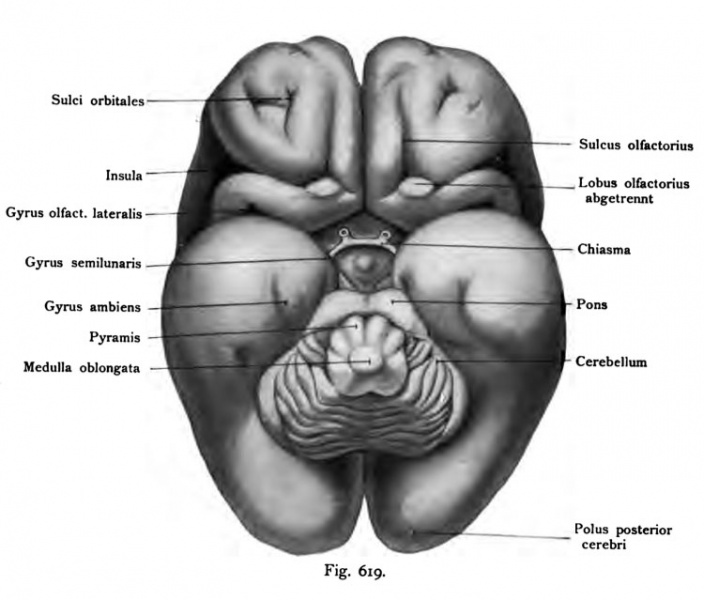
Fig. 619. Base of the brain of a human fetus from the beginning of the 6th month
(After Retzius) enlarged
The olfactory lobe is separated from tight angles to the brain pulls him forward Suicus the olfactory, lateral to the anterior edge of the island, behind which the gyrus olfactorius lateralis extends to invade after a sharp bend in the nearest to the front portion of the temporal lobe (comp. Fig. 615). Easy is the gyrus ambiens, the gyrus semilunaris seen also the optic chiasm to the optic nerve, which infuse Bulum, the following saccularis eminence, the cerebral peduncles, and medulla oblongata with cerebellum bridge. On the basal surface of the temporal and occipital no furrows are well defined at this time.
- This text is a Google translate computer generated translation and may contain many errors.
Images from - Atlas of the Development of Man (Volume 2)
(Handatlas der entwicklungsgeschichte des menschen)
- Kollmann Atlas 2: Gastrointestinal | Respiratory | Urogenital | Cardiovascular | Neural | Integumentary | Smell | Vision | Hearing | Kollmann Atlas 1 | Kollmann Atlas 2 | Julius Kollmann
- Links: Julius Kollman | Atlas Vol.1 | Atlas Vol.2 | Embryology History
| Historic Disclaimer - information about historic embryology pages |
|---|
| Pages where the terms "Historic" (textbooks, papers, people, recommendations) appear on this site, and sections within pages where this disclaimer appears, indicate that the content and scientific understanding are specific to the time of publication. This means that while some scientific descriptions are still accurate, the terminology and interpretation of the developmental mechanisms reflect the understanding at the time of original publication and those of the preceding periods, these terms, interpretations and recommendations may not reflect our current scientific understanding. (More? Embryology History | Historic Embryology Papers) |
Reference
Kollmann JKE. Atlas of the Development of Man (Handatlas der entwicklungsgeschichte des menschen). (1907) Vol.1 and Vol. 2. Jena, Gustav Fischer. (1898).
Cite this page: Hill, M.A. (2024, April 26) Embryology Kollmann619.jpg. Retrieved from https://embryology.med.unsw.edu.au/embryology/index.php/File:Kollmann619.jpg
- © Dr Mark Hill 2024, UNSW Embryology ISBN: 978 0 7334 2609 4 - UNSW CRICOS Provider Code No. 00098G
Fig. 619. Hirnbasis eines menschlichen t Fetus vom Anfang des 6. Monats.
(Nach Retzius.) Vergrößert
Der Lobus olfactorius ist dicht an dem Hirn quer abgetrennt Von ihm nach vorn zieht der Suicus olfactorius, lateral der vordere Rand der Insel, hinter dem der Gyrus olfactorius lateralis hinzieht, um nach einer scharfen Biegung in den zunächst liegenden vorderen Abschnitt des Temporallappens einzudringen (vergl. Fig. 615). Leicht ist der Gyrus ambiens, der Gyrus semi- lunaris erkennbar, ferner das Chiasma mit dem Nervus opticus, dem Infundi- bulum, der folgenden Eminentia saccularis, die Pedunculi cerebri, Brücke und Medulla oblongata mit Cerebellum. Auf der basalen Fläche des Temporal- und Occipitallappens sind noch keine Furchen um diese Zeit deutlich ausgeprägt.
File history
Click on a date/time to view the file as it appeared at that time.
| Date/Time | Thumbnail | Dimensions | User | Comment | |
|---|---|---|---|---|---|
| current | 17:13, 17 October 2011 |  | 750 × 639 (51 KB) | S8600021 (talk | contribs) | {{Kollmann1907}} Category:Human Category:Neural Fig. 619. Hirnbasis eines menschlichen t Fetus vom Anfang des 6. Monats. (Nach Retzius.) Vergrößert Der Lobus olfactorius ist dicht an dem Hirn quer abgetrennt Von ihm nach vorn zieht de |
You cannot overwrite this file.
File usage
The following page uses this file:
