File:Kollmann616.jpg
Kollmann616.jpg (496 × 567 pixels, file size: 36 KB, MIME type: image/jpeg)
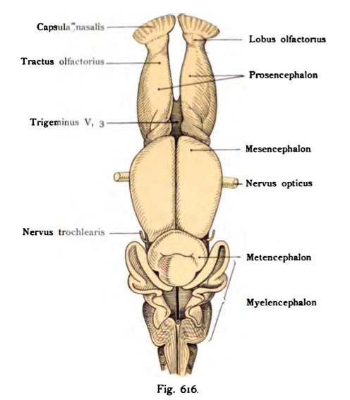
Fig. 616. Brain of a young Shark (Callorliynclius antarcticus)
Approximately the same age as that depicted his head and his skull skeleton Figure 258, 259
Here, the broad outline are shown five of the brain in a lower vertebrate. To the elongated body occur following Einzelnheitenivon front enumerated backwards pointed out: the nasal capsule to the optic tract runs olfactorius, the elongated telencephalon (telencephalon), Mes-encephalon (midbrain), the metencephalon (hindbrain), myelencephalon (After-him). The diencephalon (Zwischenhim) is not visible when viewed from above because of the strong bending of the brain tube. Nerves of the trigeminal nerve are visible bar and that the III. Branch of the optic nerve and nerve trochlear. The original 12 times magnified at Schauinsland
- This text is a Google translate computer generated translation and may contain many errors.
Images from - Atlas of the Development of Man (Volume 2)
(Handatlas der entwicklungsgeschichte des menschen)
- Kollmann Atlas 2: Gastrointestinal | Respiratory | Urogenital | Cardiovascular | Neural | Integumentary | Smell | Vision | Hearing | Kollmann Atlas 1 | Kollmann Atlas 2 | Julius Kollmann
- Links: Julius Kollman | Atlas Vol.1 | Atlas Vol.2 | Embryology History
| Historic Disclaimer - information about historic embryology pages |
|---|
| Pages where the terms "Historic" (textbooks, papers, people, recommendations) appear on this site, and sections within pages where this disclaimer appears, indicate that the content and scientific understanding are specific to the time of publication. This means that while some scientific descriptions are still accurate, the terminology and interpretation of the developmental mechanisms reflect the understanding at the time of original publication and those of the preceding periods, these terms, interpretations and recommendations may not reflect our current scientific understanding. (More? Embryology History | Historic Embryology Papers) |
Reference
Kollmann JKE. Atlas of the Development of Man (Handatlas der entwicklungsgeschichte des menschen). (1907) Vol.1 and Vol. 2. Jena, Gustav Fischer. (1898).
Cite this page: Hill, M.A. (2024, April 26) Embryology Kollmann616.jpg. Retrieved from https://embryology.med.unsw.edu.au/embryology/index.php/File:Kollmann616.jpg
- © Dr Mark Hill 2024, UNSW Embryology ISBN: 978 0 7334 2609 4 - UNSW CRICOS Provider Code No. 00098G
Fig. 616. Gehirn eines jungen Callorliynclius antarcticus
ungefähr desselben Alters wie jener, dessen Kopf und dessen Schädelgerüst abgebildet
wurde. Fig. 258, 259.
Hier soll die weitreichende Fünfgliederung des Gehirns bei einem niederen Wirbeltier gezeigt werden. An dem langgestreckten Organ treten folgende Einzelnheitenivon vorn nach hinten aufgezählt, hervor : die Nasenkapsel zu der der Tractus olfactorius zieht, das längliche Telencephalon (Endhirn), Mes- encephalon (Mittelhirn), das Metencephalon (Hinterhirn), Myelencephalon (Nach- him). Das Diencephalon (Zwischenhim) ist bei der Betrachtung von oben wegen der starken Knickung des Hirnrohres nicht zu sehen. Von Nerven sind sicht- bar der Trigeminus und zwar dessen III. Ast, der Nervus opticus und der N. trochlearis. Das Original bei Schauinsland 12 mal vergr.
File history
Click on a date/time to view the file as it appeared at that time.
| Date/Time | Thumbnail | Dimensions | User | Comment | |
|---|---|---|---|---|---|
| current | 17:04, 17 October 2011 |  | 496 × 567 (36 KB) | S8600021 (talk | contribs) | {{Kollmann1907}} Category:Neural Fig. 616. Gehirn eines jungen Callorliynclius antarcticus ungefähr desselben Alters wie jener, dessen Kopf und dessen Schädelgerüst abgebildet wurde. Fig. 258, 259. Hier soll die weitreichende Fünfglied |
You cannot overwrite this file.
File usage
The following page uses this file:

