File:Kollmann543.jpg
Kollmann543.jpg (739 × 445 pixels, file size: 63 KB, MIME type: image/jpeg)
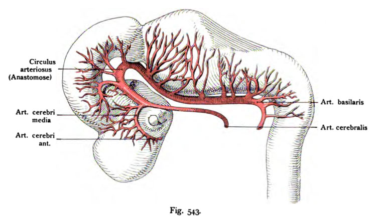
Fig. 543. Brain and its arteries in a human embryo of 9 mm
lateral view
(After Mall)
The basilar artery has a very long course through the posterior divisions of the embryonic brain. The anastomosis with the Art. cerebralis (later the circle of Willis) moves through the front section of the brain Art. cerebralis (later carotis cerebralis) sends an artery, to the orbits in the optic cup, bow, and the four brain vesicles branches provenance. Probably go out of her anterior cerebral artery and the middle cerebral artery produced. In the numerous branches of the artery basilaris, that approach the brain tube and later make significant shifts and changes.
- This text is a Google translate computer generated translation and may contain many errors.
Images from - Atlas of the Development of Man (Volume 2)
(Handatlas der entwicklungsgeschichte des menschen)
- Kollmann Atlas 2: Gastrointestinal | Respiratory | Urogenital | Cardiovascular | Neural | Integumentary | Smell | Vision | Hearing | Kollmann Atlas 1 | Kollmann Atlas 2 | Julius Kollmann
- Links: Julius Kollman | Atlas Vol.1 | Atlas Vol.2 | Embryology History
| Historic Disclaimer - information about historic embryology pages |
|---|
| Pages where the terms "Historic" (textbooks, papers, people, recommendations) appear on this site, and sections within pages where this disclaimer appears, indicate that the content and scientific understanding are specific to the time of publication. This means that while some scientific descriptions are still accurate, the terminology and interpretation of the developmental mechanisms reflect the understanding at the time of original publication and those of the preceding periods, these terms, interpretations and recommendations may not reflect our current scientific understanding. (More? Embryology History | Historic Embryology Papers) |
Reference
Kollmann JKE. Atlas of the Development of Man (Handatlas der entwicklungsgeschichte des menschen). (1907) Vol.1 and Vol. 2. Jena, Gustav Fischer. (1898).
Cite this page: Hill, M.A. (2024, April 26) Embryology Kollmann543.jpg. Retrieved from https://embryology.med.unsw.edu.au/embryology/index.php/File:Kollmann543.jpg
- © Dr Mark Hill 2024, UNSW Embryology ISBN: 978 0 7334 2609 4 - UNSW CRICOS Provider Code No. 00098G
Fig. 543. Gehirn und seine Arterien bei einem menschlichen Embryo von
9 mm Scheitelsteifilänge.
(Nach Mall.)
Die Arteria basilaris hat einen sehr langen Verlauf durch die hintere Abteilung des embryonalen Gehirns. Die Anastomose mit der Art. cerebralis
(später Circulus arteriosus Willisii) zieht durch den vorderen Abschnitt des Ge-
hirns. Die Art. cerebralis (später Carotis cerebralis) entsendet eine Arterie,
die im Bogen den Augenbecher umkreist, und an das Grofahirnbläschen vier
Äste abgibt. Aus ihr gehen wahrscheinlich die Arteria cerebri anterior und
die Arteria cerebri media hervor. In den zahlreichen Ästen der Arteria basi-
laris, die mit großer Regelmäßigkeit an das Hirnrohr herantreten, erfolgen
später beträchtliche Verschiebungen und Abänderungen.
File history
Click on a date/time to view the file as it appeared at that time.
| Date/Time | Thumbnail | Dimensions | User | Comment | |
|---|---|---|---|---|---|
| current | 00:12, 17 October 2011 |  | 739 × 445 (63 KB) | S8600021 (talk | contribs) | {{Kollmann1907}} |
You cannot overwrite this file.
File usage
The following page uses this file:

