File:Kollmann541.jpg
Kollmann541.jpg (757 × 535 pixels, file size: 74 KB, MIME type: image/jpeg)
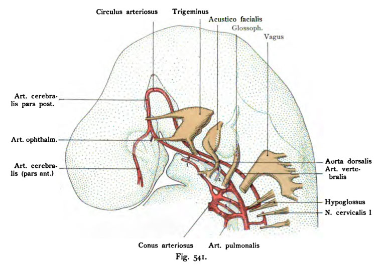
Fig. 541. The aortic arch of a human embryo of 7 mm CRL
(According to Tandler.)
It is only the left half of the aortic arch represented, just as seen from the left side of the head. This embryo has six aortic arches. The fifth draws from the pulmonary trunk aorta arch. The connection tion of the ventral aorta with the dorsal aorta in the area of the first arch interrupted. This reduction has already occurred, but the course is drawn. The second arc is in a state of regression. Between the ventral aorta and the third (carotid arch) is an island formation. The foregut with its pharyngeal pouches is still similar to the embryo of 5 mm (Fig. 540).
- This text is a Google translate computer generated translation and may contain many errors.
Images from - Atlas of the Development of Man (Volume 2)
(Handatlas der entwicklungsgeschichte des menschen)
- Kollmann Atlas 2: Gastrointestinal | Respiratory | Urogenital | Cardiovascular | Neural | Integumentary | Smell | Vision | Hearing | Kollmann Atlas 1 | Kollmann Atlas 2 | Julius Kollmann
- Links: Julius Kollman | Atlas Vol.1 | Atlas Vol.2 | Embryology History
| Historic Disclaimer - information about historic embryology pages |
|---|
| Pages where the terms "Historic" (textbooks, papers, people, recommendations) appear on this site, and sections within pages where this disclaimer appears, indicate that the content and scientific understanding are specific to the time of publication. This means that while some scientific descriptions are still accurate, the terminology and interpretation of the developmental mechanisms reflect the understanding at the time of original publication and those of the preceding periods, these terms, interpretations and recommendations may not reflect our current scientific understanding. (More? Embryology History | Historic Embryology Papers) |
Reference
Kollmann JKE. Atlas of the Development of Man (Handatlas der entwicklungsgeschichte des menschen). (1907) Vol.1 and Vol. 2. Jena, Gustav Fischer. (1898).
Cite this page: Hill, M.A. (2024, April 27) Embryology Kollmann541.jpg. Retrieved from https://embryology.med.unsw.edu.au/embryology/index.php/File:Kollmann541.jpg
- © Dr Mark Hill 2024, UNSW Embryology ISBN: 978 0 7334 2609 4 - UNSW CRICOS Provider Code No. 00098G
Fig. 541. Die Aortenbogen eines menschlichen Embryo von 7 mm Nacken-
steifilänge.
(Nach Tandler.)
Es ist nur die linke Hälfte der Aortenbogen dargestellt, wie denn auch der Kopf von der linken Seite gesehen ist Dieser Embryo hat sechs Aorten- bogen. Der fünfte zieht vom Aortentruncus zum Pulmonalisbogen. Die Verbin- dung der Aorta ventralis mit der Aorta dorsalis ist im Bereich des ersten Bogens unterbrochen. Hier ist schon Reduktion eingetreten, doch ist der Verlauf ein- gezeichnet. Der zweite Bogen befindet sich im Stadium der Rückbildung. Zwischen der Aorta ventralis und dem dritten (Carotisbogen) besteht eine Inselbildung. Der Kopfdarm mit seinen Schlundtaschen ist noch ähnlich wie bei dem Embryo von 5 mm (Fig. 540).
File history
Click on a date/time to view the file as it appeared at that time.
| Date/Time | Thumbnail | Dimensions | User | Comment | |
|---|---|---|---|---|---|
| current | 00:11, 17 October 2011 |  | 757 × 535 (74 KB) | S8600021 (talk | contribs) | {{Kollmann1907}} |
You cannot overwrite this file.
File usage
The following page uses this file:

