File:Kollmann540.jpg
Kollmann540.jpg (746 × 376 pixels, file size: 62 KB, MIME type: image/jpeg)
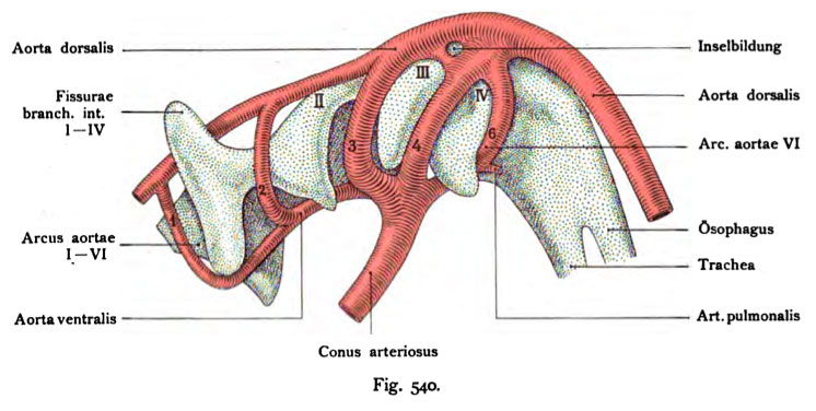
Fig. 540. Aortic arch of a human embryo of 5 mm greatest length
including the pharynx and the inner gill-pouches.
(75 times enlarged.)
(According to Tandler.)
There are four pharyngeal pouches formed. Towering over the first beyond the pharynx. The conus arteriosus is still simple. There are five Aortic arch to see: the I, II, III, IV and VI. The sixth bypasses inner fourth pharyngeal pouch caudally and medially. He dismisses a small Branch to the tracheal system. At the mouth of the 6th aortic arch is an islet formation. The rising of the ventral pharyngeal part of the conus arteriosus, from which spring from the I. and II arch means Ventral aorta, the longitudinal strain, occur in the aortic arch is, Dorsal aorta. The third aortic arch is also called carotid arteries, the known as pulmonary sixth arch.
- This text is a Google translate computer generated translation and may contain many errors.
Images from - Atlas of the Development of Man (Volume 2)
(Handatlas der entwicklungsgeschichte des menschen)
- Kollmann Atlas 2: Gastrointestinal | Respiratory | Urogenital | Cardiovascular | Neural | Integumentary | Smell | Vision | Hearing | Kollmann Atlas 1 | Kollmann Atlas 2 | Julius Kollmann
- Links: Julius Kollman | Atlas Vol.1 | Atlas Vol.2 | Embryology History
| Historic Disclaimer - information about historic embryology pages |
|---|
| Pages where the terms "Historic" (textbooks, papers, people, recommendations) appear on this site, and sections within pages where this disclaimer appears, indicate that the content and scientific understanding are specific to the time of publication. This means that while some scientific descriptions are still accurate, the terminology and interpretation of the developmental mechanisms reflect the understanding at the time of original publication and those of the preceding periods, these terms, interpretations and recommendations may not reflect our current scientific understanding. (More? Embryology History | Historic Embryology Papers) |
Reference
Kollmann JKE. Atlas of the Development of Man (Handatlas der entwicklungsgeschichte des menschen). (1907) Vol.1 and Vol. 2. Jena, Gustav Fischer. (1898).
Cite this page: Hill, M.A. (2024, April 26) Embryology Kollmann540.jpg. Retrieved from https://embryology.med.unsw.edu.au/embryology/index.php/File:Kollmann540.jpg
- © Dr Mark Hill 2024, UNSW Embryology ISBN: 978 0 7334 2609 4 - UNSW CRICOS Provider Code No. 00098G
Fig. 540. Aortenbogen eines menschlichen Embryo von 5 mm gröfiter Länge
samt dem Pharynx und den inneren Kiementaschen.
(75 mal vergrößert.) (Nach Tandler.)
Es sind 4 innere Schlundtaschen ausgebildet. Die erste ragt hoch über den Pharynx hinaus. Der Conus arteriosus ist noch einfach. Es sind fünf Aortenbogen zu sehen: der L, IL, III., IV. und VI. Der sechste umgeht die vierte innere Schlundtasche kaudal-und medialwärts. Er entläßt einen kleinen Ast zur Trachealanlage. An der Mündungsstelle des 6. Aortenbogens findet sich eine kleine Inselbildung. Der an der ventralen Pharynxwand aufsteigende Teil des Conus arteriosus, aus dem der I. und IL Bogen entspringen, heißt Aorta ventralis; der Längsstamm, in den die Aortenbogen eintreten, heißt Aorta dorsalis. Der dritte Aortenbogen wird auch als Carotidenbogen , der sechste als Pulmonalisbogen bezeichnet.
File history
Click on a date/time to view the file as it appeared at that time.
| Date/Time | Thumbnail | Dimensions | User | Comment | |
|---|---|---|---|---|---|
| current | 00:11, 17 October 2011 |  | 746 × 376 (62 KB) | S8600021 (talk | contribs) | {{Kollmann1907}} |
You cannot overwrite this file.
File usage
The following page uses this file:

