File:Kollmann535.jpg
Kollmann535.jpg (646 × 577 pixels, file size: 81 KB, MIME type: image/jpeg)
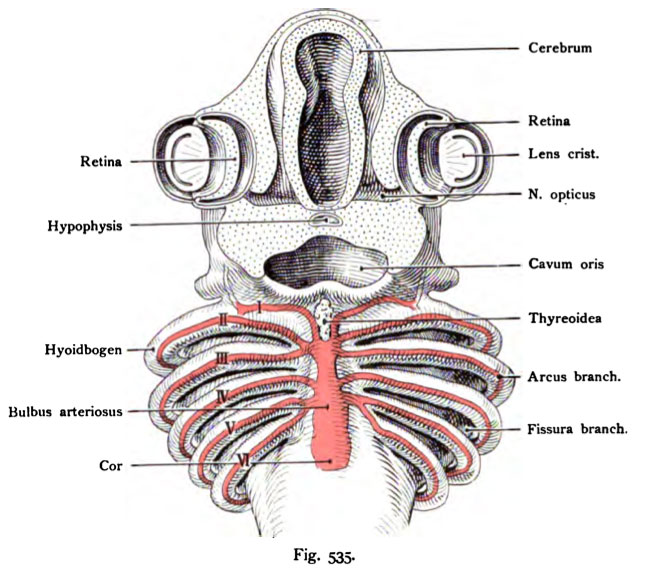
Fig. 535. The aortic arch in the shark embryo (Pristiurus)
Where the original relations of the aortic arch to the gills still exist.
Seen from below.
(Partly to Dohrn.)
The forehead is removed by a frontal section. The heart is away, on the ventral wall of the foregut is the truncus arterioles and divided, with most selachians, in 6 aortic arch on each side, the
Foregut enclose an arc to lead into dorsal to the aorta. Much of this behavior of the truncus arteriosus versa in mammals and humans again. (See Figures 537 and 538)
- This text is a Google translate computer generated translation and may contain many errors.
Images from - Atlas of the Development of Man (Volume 2)
(Handatlas der entwicklungsgeschichte des menschen)
- Kollmann Atlas 2: Gastrointestinal | Respiratory | Urogenital | Cardiovascular | Neural | Integumentary | Smell | Vision | Hearing | Kollmann Atlas 1 | Kollmann Atlas 2 | Julius Kollmann
- Links: Julius Kollman | Atlas Vol.1 | Atlas Vol.2 | Embryology History
| Historic Disclaimer - information about historic embryology pages |
|---|
| Pages where the terms "Historic" (textbooks, papers, people, recommendations) appear on this site, and sections within pages where this disclaimer appears, indicate that the content and scientific understanding are specific to the time of publication. This means that while some scientific descriptions are still accurate, the terminology and interpretation of the developmental mechanisms reflect the understanding at the time of original publication and those of the preceding periods, these terms, interpretations and recommendations may not reflect our current scientific understanding. (More? Embryology History | Historic Embryology Papers) |
Reference
Kollmann JKE. Atlas of the Development of Man (Handatlas der entwicklungsgeschichte des menschen). (1907) Vol.1 and Vol. 2. Jena, Gustav Fischer. (1898).
Cite this page: Hill, M.A. (2024, April 27) Embryology Kollmann535.jpg. Retrieved from https://embryology.med.unsw.edu.au/embryology/index.php/File:Kollmann535.jpg
- © Dr Mark Hill 2024, UNSW Embryology ISBN: 978 0 7334 2609 4 - UNSW CRICOS Provider Code No. 00098G
Fig. 535. Die Aortenbogen eines Haifischembryo (Pristiurus)
bei dem die ursprünglichen Beziehungen der Aortenbogen zu den Kiemenbogen
noch bestehen. Etwas von unten gesehen.
(Teilweise nach Do hm.)
Der Vorderkopf ist durch einen frontalen Schnitt abgetragen. Das Herz ist entfernt, auf der ventralen Wand des Kopfdarms liegt der Truncus arteriosus und teilt sich, bei den meisten Selachiern, in 6 Aortenbogen für jede Seite, die den Kopfdarm bogenförmig umgreifen, um in die Aorta dorsalis einzumünden. Ein großer Teil dieses Verhaltens des Truncus arteriosus kehrt bei den Säugetieren und dem Menschen wieder. (Siehe die Fig. 537 und 538.)
File history
Click on a date/time to view the file as it appeared at that time.
| Date/Time | Thumbnail | Dimensions | User | Comment | |
|---|---|---|---|---|---|
| current | 00:09, 17 October 2011 |  | 646 × 577 (81 KB) | S8600021 (talk | contribs) | {{Kollmann1907}} |
You cannot overwrite this file.
File usage
The following 3 pages use this file:

