File:Keibel1910 plate03.jpg
Original file (1,891 × 2,476 pixels, file size: 425 KB, MIME type: image/jpeg)
Plate 3. Development of the Salamander
The series of eggs, embryos and larvae of Necturus, from which the following descriptions and the appended illustrations were made, were collected May I5th, 1903 and kept at a water temperature of 17o — 18o C. The illustrations are copied from the original water colored pictures which were made by Mr. Leonard H. Wilder, under the direction of the senior author. It should be emphasized that the ages, measurements and illustrations are all made from the living objects.
Note - Magnifications refer to original print versions.
Fig. 25. (X 5.)
Side view of embryo 23 days 10 hrs. old, length 9 mm, 20 — 22 pairs of myotomes. General outline of the body straighter. Head free from yolk. Caudal enlargement becoming free. Optic vesicles and forebrain much larger. Mandibular, hyoid, first branchial, and common anläge of second and third branchial arches well defined. Otic vesicle visible above hyoid arch.
Fig. 26. (X 5.)
Side view of embryo 24 days 22 hrs. old, length 10 mm, 23 — 24 pairs of myotomes. General outline of body of embryo straighter, less curved laterally. Head and caudal extremities free from yolk. Yolk becoming oval. Optic vesicles prominent. Ear better defined. Olfactory pits present. The mandibular, liyoid and first branchial arches are distinct. The second and third branchial arches are not yet differentiated, a slight process on the first branchial indicates the beginning of the gill bar. The anläge of the heart is visible just beneath the arches.
Fig. 27. (X 5.)
Side view of embryo 26 days old , length 1 1 mm , 26—27 myotomes. General outline of body straighter than in preceding stage. Head projects some 3 mm beyond margin of yolk; tail projects 1.2 mm, is thinner laterally but broader dorso-ventrally. Eye, ear, nasal pits and mouth well defined. Maxillary process discernible. Mandibular arches longer, but ventral ends widely separated. Second and third branchial arches formed. Gill bars present on three branchial arches. Anterior limb buds indicated; faint anläge of posterior limb buds. Yolk pear-shaped. Heart prominent. First surface capillaries present although not indicated in figure.
Fig. 28. (X 5)
Side view of embryo 30 days 8 hrs. old, length 13 mm, 30 — 31 myotomes. The trunk of the embryo is nearly straight. At level of the posterior gill there is a pronounced neck bend and at the level of the posterior limbs a striking downward bend of the tail. The epiphysis shows in surface views. The lens is discernible. The ear is still vi.sible. The external nasal openings are sharply defined. The boundaries of the mouth are better outlined owing to the approximation of the ventral ends of the mandibular arches. The hyoid arch is becoming obscured. The gill bars are prominent on the three branchial arches. The anterior limb buds project dorsally about .5 mm above the surface of the bod3\ The posterior limb buds are but slight elevations. The yolk is pear-shaped with its dorsal surface much flattened. The auricular and ventricular portions of the heart are apparent. The surface of the yolk is covered by a dense network of capillaries which for the most part convey blood antero-ventrally to the abdominal vein. Considerable pigment is present in the trunk region although but little has reached the outer portion of the dermis.
Fig. 29. (X 5)
Side view of embryo 36 days 16 hrs. old, length 16 mm, 36-38 myotomes. In general outline the embryo shows a numljcr of striking changes. The neck bend is not so pronounced. The tail bend is scarcely noticeable. There is a striking increase in dorso-ventral width of tail. The cerebral hemispheres are well defined. The eye is now prominent and the lens better defined. The ear is no longer visible in surface views. The mouth is well defined. The ends of the mandibular arches are closely approximated but not united. The hyoid and branchial arches are more obscure. Anlagen of gill fiiaments present on gill bars. Anterior limbs project dorsally. Posterior limbs are short ridges extending in horizontal plane. The yolk is elongated and reduced in diameter both dorso-ventrally and laterally. Surface blood vessels as in preceding stage, excepting that they are now apparent in the gill bars. The chromatophores are most numerous in the anterior and dorsal portions of the head.
Fig. 30. (X 5.)
Side view of einhryo 40 days 20 hrs. old, length 18 mm, 44—46 myotomes. The outline of the body shows a marked ventral curvature of the trunk, less pronounced neck bend, and further increase in the dorso-ventral width of the tail. The eye is very prominent owing to the pigment in the retina. Ear not visible externally. Nasal openings very small. The mandibular arches have coalesced. The boundaries of the other arches are no longer discernible. Gill filaments well developed. Anterior limbs about i mm long project dorso-posteriorly. The yolk is elongated oval. Abdominal vein and branchial blood vessels prominent. Pigment present in dorsal portion of head, also along dorsal and lateral portions of trunk and tail. The yolk is unpigmented excepting along dorsal margin.
Fig- 31. (X 5.)
Side view of larva 49 days old, lergth 21 mm. General outline of body decidedly different. Head bend obliterated, slight upward curve in trunk. Tail broader. Eye more deeply pigmented. Gill bars very long, extending to level of end of anterior limb. From three to five lateral filaments on each gill bar. Anterior limbs project postero-ventrally ; three digits formed. Posterior limbs directed caudad ; no trace of digits. Yolk much elongated. Network of capillaries denser. Large lateral arteries, at level of upper margin of yolk, very prominent. Well defined longitudinal bands of pigment.
Fig: 32. (X 5.)
Side view of larva 61 days old, length 25 mm. General outline of body shows less dorsal curvature of trunk. Tail much longer in proportion to length of trunk and much broader dorso-ventrally. Gill bars longer, each posessing six to eight lateral filaments. Anterior and posterior limbs directed postero-ventrally. Anterior 3 mm long, posterior 2 mm long. Each limb shows four digits. The distribution of pigment is essentially similar to that observed in the 21 mm larva, the bands however are more sharply defined. Chromatophores in the gill bars and limbs and beginning to extend over the dorsal surface of the yolk.
Fig. 33. (X 5.)
Side view of larva 70 days 4 hrs. old, length 28 mm. The general outline of the body is slenderer than at any time preceding. The rapid absorption of the yolk has brought its ventral surface nearly to the level of the ventral surfaces of the head and tail. The gill bars curve dorsally and possess fi;om ten to twelve pairs of lateral filaments. The tail is somewhat constricted at the level of the posterior limbs. The limbs and digits are better developed and are now used in locomotion. Pigmentation is denser than in 25 mm larva, but same general arrangement of bands prevails.
Fig. 34. (X 5.)
Side view of larva 97 days old, length 34 mm. In general outline the larva begins to resemble the adult. The yolk is well absorbed. The tail is very broad and now used as a powerful caudal fin in swimming. The gill bars project dorsally and have a large number of filaments. The legs project far below the ventral surface of the body. In color the same general pattern prevails as in the 28 mm larva. There are some minor changes, the Hght band is broader and better defined, and extensions of pigment over the yolk have been so uneven that a number of irregulär oval areas are left unpigmented, causing a mottled appearance in this region.
Fig. 35. (X 5.)
Side view of larva 126 days old, length 39 mm. The young Nedurus now conforms in outline to the adult. In color hovvever it is decidedly different.
Fig. 25-27.
Fig. 28-29.
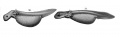
Fig. 30-31.
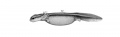
Fig. 32.
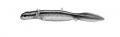
Fig. 33.
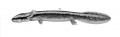
Fig. 34.
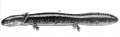
Fig. 35.
| Historic Disclaimer - information about historic embryology pages |
|---|
 |
Reference
Eycleshymer AC. and Wilson JM. Normal Plates of the Development of the Salamander Embryo (Nectürüs maculosus). Vol. 11 in series by Keibel F. Normal plates of the development of vertebrates (Normentafeln zur Entwicklungsgeschichte der Wirbelthiere) Fisher, Jena., Germany.
Cite this page: Hill, M.A. (2024, April 26) Embryology Keibel1910 plate03.jpg. Retrieved from https://embryology.med.unsw.edu.au/embryology/index.php/File:Keibel1910_plate03.jpg
- © Dr Mark Hill 2024, UNSW Embryology ISBN: 978 0 7334 2609 4 - UNSW CRICOS Provider Code No. 00098G
File history
Click on a date/time to view the file as it appeared at that time.
| Date/Time | Thumbnail | Dimensions | User | Comment | |
|---|---|---|---|---|---|
| current | 12:52, 10 January 2015 | | 1,891 × 2,476 (425 KB) | Z8600021 (talk | contribs) |
You cannot overwrite this file.
File usage
The following page uses this file: