File:Keibel1910 plate02.jpg

Original file (1,891 × 2,476 pixels, file size: 360 KB, MIME type: image/jpeg)
Plate 2. Development of the Salamander
The series of eggs, embryos and larvae of Necturus, from which the following descriptions and the appended illustrations were made, were collected May I5th, 1903 and kept at a water temperature of 17o — 18o C. The illustrations are copied from the original water colored pictures which were made by Mr. Leonard H. Wilder, under the direction of the senior author. It should be emphasized that the ages, measurements and illustrations are all made from the living objects.
Note - Magnifications refer to original print versions.
Fig. 13. (X 10.)
Top view of egg 13 days 3 hrs. old. Small circular blastopore. Embryonic anläge triangulär in outline ; lateral boundaries indistinct. First appearance of neural groove. Roof of segmentation cavity thinner, making its boundaries distinct.
Fig. 14. (X 10.)
Top view of egg 14 days 4 hrs. old. Blastopore smaller, lateral margins of anterior portion of embryo bounded by short broad ridges which are the beginnings of the lateral portions of the neural fold. At anterior margin of embryo there is a transverse crescentic ridge which is beginning of transverse portion of neural fold. Neural groove deep but does not extend either to transverse portion of neural fold or to blastopore. Segmentation cavity crescentic.
Fig. 15. (X 10.)
Top view of egg 14 days 19 hrs. old. Blastopore much reduced, circular. The yolk plug is not visible in this egg. Lateral and transverse portions of neural fold united to form continuous fold around anterior portion of embryo. Lateral boundaries of posterior portion of embryo not defined. Neural groove not as long, nor as distinct as in preceding stage. Dark crescentic area in front of embryo is segmentation cavity.
Fig. 16. (X 10.)
Top view of egg 15 days 15 hrs. old. Blastopore small, circular; yolk plug visible. Neural fold prominent, its free ends extend nearly to blastopore. Neural groove deep and narrow at anterior end, broad and shallow at posterior end, fades out just in front of blastopore. A part of the segmentation cavity is still apparent in front of the embryo.
Fig. 17. (X 10.)
Top view of egg 16 days 6 hrs. old. Blastopore reduced to a very minute circular aperture. Neural plate narrower than in preceding stage. Neural fold prominent, its free ends coalescing at blastopore. Neural groove extends to transverse portion of fold but does not reach blastopore. Segmentation cavity no longer visible in surface views.
Fig. 18. (X 10.)
Top view of embryo 17 days 2 hrs. old. Blastopore an elongated narrow aperture between ends of neural fold. Neural plate narrower than in preceding stage. The constricted portion represents in a general way the division between head and trunk. Neural fold most prominent in head region.
Fig. 19. (X 10.)
Top view of egg 17 days 17 hrs. old. Blastopore no longer visible. Neural plate narrowest posteriorly; broad in head region, showing boundary zone between head and trunk. Lateral portions of fold coalesced at posterior end of embryo. At anterior end of embryo a deep groove partially separates the two halves of the neural fold.
Fig. 20. (X 10.)
Top view of egg 18 days 13 hrs. old. Lateral portions of neural fold almost united except in head region where they are still widely separated. In the antero-lateral portions of the fold are slight evaginations which are the beginnings of the optic vesicles.
Fig. 21. (X 10.)
Top view of egg 18 days 15 hrs. old, 3 or 4 pairs of myotomes. Lateral portions of neural fold widely separated in head region, more closely approximated in anterior trunk region, coalesced in tail.
Fig. 22. (X 10.) Dorso-lateral view of embryo 20 days 10 hrs. old, length 6 mm, 6 pairs of myotomes. Outline of body conforms to curvature of egg. Head end of embryo shows three longitudinal ridges; middle ridge lies slightly above level of lateral ridges. The middle one is common anläge of fore, mid and bind brain. The lateral ones are the common anläge of the optic vesicles and branchial arches. Anus formed.
Fig. 23. (X 10.)
Side view of embryo 21 days 2 hrs. old, length 7 mm, 10 — 12 pairs of myotomes. General outline of body conforms to curvature of egg. Head slightly raised above surface of yolk. Slight enlargement at end of tail. A distinct enlargement of anterior end of head shows optic vesicles; just posterior to this enlargement is the anläge of the branchial arches. Anus shows just below tip of tail.
Fig. 24. (X 10.)
Dorso-lateral view of embryo 22 days 17 hrs. old, length 8 mm, l6~18 pairs of myotomes. Embryo much curved laterally. Anterior half of head free from yolk. Caudal enlargement more prominent. Optic vesicles and mandibular arch well defined. The hyoid and first branchial arches are discernible ; also the common anläge of the second and third branchial arches.
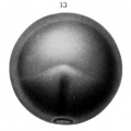
Fig. 13.
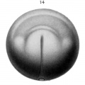
Fig. 14.
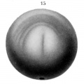
Fig. 15.
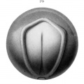
Fig. 16.
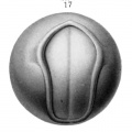
Fig. 17.
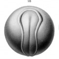
Fig. 18.
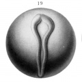
Fig. 19.
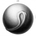
Fig. 20.
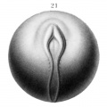
Fig. 21.
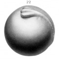
Fig. 22.
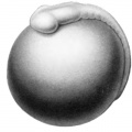
Fig. 23.
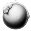
Fig. 24.


| Historic Disclaimer - information about historic embryology pages |
|---|
 |
Reference
Eycleshymer AC. and Wilson JM. Normal Plates of the Development of the Salamander Embryo (Nectürüs maculosus). Vol. 11 in series by Keibel F. Normal plates of the development of vertebrates (Normentafeln zur Entwicklungsgeschichte der Wirbelthiere) Fisher, Jena., Germany.
Cite this page: Hill, M.A. (2024, April 25) Embryology Keibel1910 plate02.jpg. Retrieved from https://embryology.med.unsw.edu.au/embryology/index.php/File:Keibel1910_plate02.jpg
- © Dr Mark Hill 2024, UNSW Embryology ISBN: 978 0 7334 2609 4 - UNSW CRICOS Provider Code No. 00098G
File history
Click on a date/time to view the file as it appeared at that time.
| Date/Time | Thumbnail | Dimensions | User | Comment | |
|---|---|---|---|---|---|
| current | 12:52, 10 January 2015 | | 1,891 × 2,476 (360 KB) | Z8600021 (talk | contribs) |
You cannot overwrite this file.
File usage
There are no pages that use this file.