File:Keibel1910 plate01.jpg

Original file (1,891 × 2,476 pixels, file size: 444 KB, MIME type: image/jpeg)
Plate 1. Development of the Salamander
The series of eggs, embryos and larvae of Necturus, from which the following descriptions and the appended illustrations were made, were collected May I5th, 1903 and kept at a water temperature of 17o — 18o C. The illustrations are copied from the original water colored pictures which were made by Mr. Leonard H. Wilder, under the direction of the senior author. It should be emphasized that the ages, measurements and illustrations are all made from the living objects.
Note - Magnifications refer to original print versions.
Fig. I. (X 10.)
Side view of egg 1 day 4 hrs. after deposition. The first cleavage groove has reached the lower pole of the egg. Second grooves extend to level of the equator of the egg.
Fig. 2. (X 10.)
Side view of egg 1 day 8 hrs. after deposition. The second cleavage grooves have reached the equator. The grooves of the third cleavage pass in meridional planes, but have not yet reached the equator.
Fig. 3. (X 10.)
Side view of egg 1 day 12 hrs. old. Five cleavage grooves have reached lower pole, dividing lower hemisphere into six segments.
Fig. 4. (X 10.)
Side view of egg 1 day 16 hrs, old. The greater number of cleavage grooves pass in meridional planes, many are latitudinal and some nearly radial. The upper surface of the egg shows sixteen segments, the lower nine.
Fig. 5. (X 10.)
Side view of egg 1 day 20 hrs. old. The upper surface of the egg shows some fifty segments, the lower nine.
Fig. 6. (X 10.)
Side view of egg 2 days 2 hrs. old. The upper surface of the egg shows more than one hundred segments, the lower twelve.
Fig. 7. (X 10.)
Side view of egg 2 days 7 hrs. old. The upper surface of egg shows about two hundred cells. The lower portion is in about same stage as described in Fig. 6.
Fig. 8. (X lo.)
Side view of egg 2 days 12 hrs. old. The upper surface of egg shows some five hundred cells, the lower about forty.
Fig. 9. (X lo.)
Top view of egg 4 days 4 hrs. old. Segmentation cavity shows through thin translucent roof. Blastopore not präsent.
Fig. 10. (X 10.)
Bottom view of egg 6 days 16 hrs. old. Crescentic blastopore. Line of invagination sharply separates large yolk cells from small cells of blastodisc.
Fig. 11. (X 10.)
Dorso-lateral view of egg 10 days 10 hrs. old. Large circular blastopore; faint indication of embryonic anläge.
Fig. 12. (X 10.)
Side view of egg 10 days 16 hrs. old. Large circular blastopore. Anlage of mesial portion of embryo above dorsal lip of blastopore. Segmentation cavity faintly outlined.
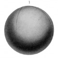
Fig. 1. 1 day 4 hrs.
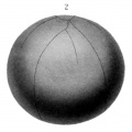
Fig. 2.
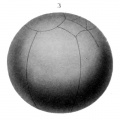
Fig. 3.
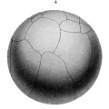
Fig. 4.
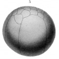
Fig. 5.
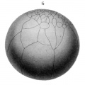
Fig. 6.
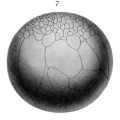
Fig. 7.
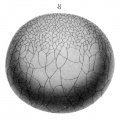
Fig. 8. 2 days 12 hrs.
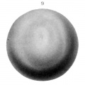
Fig. 9. 4 days 4 hrs. old.
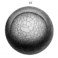
Fig. 10. 6 days 16 hrs. old.
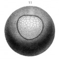
Fig. 11. 10 days 10 hrs.
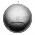
Fig. 12. 10 days 16 hrs.


| Historic Disclaimer - information about historic embryology pages |
|---|
 |
Reference
Eycleshymer AC. and Wilson JM. Normal Plates of the Development of the Salamander Embryo (Nectürüs maculosus). Vol. 11 in series by Keibel F. Normal plates of the development of vertebrates (Normentafeln zur Entwicklungsgeschichte der Wirbelthiere) Fisher, Jena., Germany.
Cite this page: Hill, M.A. (2024, April 26) Embryology Keibel1910 plate01.jpg. Retrieved from https://embryology.med.unsw.edu.au/embryology/index.php/File:Keibel1910_plate01.jpg
- © Dr Mark Hill 2024, UNSW Embryology ISBN: 978 0 7334 2609 4 - UNSW CRICOS Provider Code No. 00098G
File history
Click on a date/time to view the file as it appeared at that time.
| Date/Time | Thumbnail | Dimensions | User | Comment | |
|---|---|---|---|---|---|
| current | 12:52, 10 January 2015 | | 1,891 × 2,476 (444 KB) | Z8600021 (talk | contribs) |
You cannot overwrite this file.
File usage
The following page uses this file: