File:Hughes1950 fig01.jpg

Original file (1,280 × 730 pixels, file size: 140 KB, MIME type: image/jpeg)
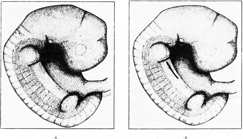
Fig. 1. Human Embryo 3.5 to 6 mm CRL
In the very earliest embryos examined, 3-5 mm., 4-5 mm., 5-5 mm. and 6-0 mm., there were no signs of a developing nipple. In the latter three, the ectoderm surrounding the origins of the limb buds, and on the lateral walls of the trunk between them, displayed a mild differentiation into two layers of cells. This ectoderm has been referred to as the “ mammary band ” (Fig. 1 (a) ).
(A) The “ mammary band ” (darkened area) surrounds the limb buds and extends between them on the lateral wall of the trunk. (B) The mammary crest appealrs wilthin the mammary band area and is represented here as a b ack ine.
Reference
Hughes ES. Development of the mammary gland. (1950) Ann R Coll Surg Engl. 6(2):99-119. PMID 19309885
Cite this page: Hill, M.A. (2024, April 27) Embryology Hughes1950 fig01.jpg. Retrieved from https://embryology.med.unsw.edu.au/embryology/index.php/File:Hughes1950_fig01.jpg
- © Dr Mark Hill 2024, UNSW Embryology ISBN: 978 0 7334 2609 4 - UNSW CRICOS Provider Code No. 00098G
File history
Click on a date/time to view the file as it appeared at that time.
| Date/Time | Thumbnail | Dimensions | User | Comment | |
|---|---|---|---|---|---|
| current | 10:07, 15 August 2018 |  | 1,280 × 730 (140 KB) | Z8600021 (talk | contribs) | |
| 10:05, 15 August 2018 |  | 2,005 × 1,302 (337 KB) | Z8600021 (talk | contribs) | {{Ref-Hughes1950}} |
You cannot overwrite this file.
File usage
The following page uses this file: