File:Gray1013.jpg
Gray1013.jpg (562 × 500 pixels, file size: 56 KB, MIME type: image/jpeg)
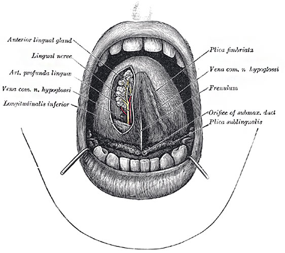
Fig. 1013. The Mouth Cavity
The apex of the tongue is turned upward, and on the right side a superficial dissection of its under surface has been made.
The tongue (lingua) is the principal organ of the sense of taste, and an important organ of speech; it also assists in the mastication and deglutition of the food. It is situated in the floor of the mouth, within the curve of the body of the mandible.
The tongue inferior surface (facies inferior linguæ under surface) (Fig. 1013) is connected with the mandible by the Genioglossi; the mucous membrane is reflected from it to the lingual surface of the gum and on to the floor of the mouth, where, in the middle line, it is elevated into a distinct vertical fold, the frenulum linguæ. On either side lateral to the frenulum is a slight fold of the mucous membrane, the plica fimbriata, the free edge of which occasionally exhibits a series of fringe-like processes. The apex of the tongue, part of the inferior surface, the sides, and dorsum are free.
Glands of the Tongue
- Mucous glands are similar in structure to the labial and buccal glands. They are found especially at the back part behind the vallate papillæ, but are also present at the apex and marginal parts. In this connection the anterior lingual glands (Blandin or Nuhn) require special notice. They are situated on the under surface of the apex of the tongue (Fig. 1013), one on either side of the frenulum, where they are covered by a fasciculus of muscular fibers derived from the Styloglossus and Longitudinalis inferior. They are from 12 to 25 mm. long, and about 8 mm. broad, and each opens by three or four ducts on the under surface of the apex.
- Serous glands occur only at the back of the tongue in the neighborhood of the taste-buds, their ducts opening for the most part into the fossæ of the vallate papillæ. These glands are racemose (clustered), the duct of each branching into several minute ducts, which end in alveoli, lined by a single layer of more or less columnar epithelium. Their secretion is of a watery nature, and probably assists in the distribution of the substance to be tasted over the taste area. (Ebner.)
- Links: Tongue Development | Salivary Gland Development | Head Development | Musculoskeletal System Development |
- Gray's Images: Development | Lymphatic | Neural | Vision | Hearing | Somatosensory | Integumentary | Respiratory | Gastrointestinal | Urogenital | Endocrine | Surface Anatomy | iBook | Historic Disclaimer
| Historic Disclaimer - information about historic embryology pages |
|---|
| Pages where the terms "Historic" (textbooks, papers, people, recommendations) appear on this site, and sections within pages where this disclaimer appears, indicate that the content and scientific understanding are specific to the time of publication. This means that while some scientific descriptions are still accurate, the terminology and interpretation of the developmental mechanisms reflect the understanding at the time of original publication and those of the preceding periods, these terms, interpretations and recommendations may not reflect our current scientific understanding. (More? Embryology History | Historic Embryology Papers) |
| iBook - Gray's Embryology | |
|---|---|

|
|
Reference
Gray H. Anatomy of the human body. (1918) Philadelphia: Lea & Febiger.
Cite this page: Hill, M.A. (2024, April 26) Embryology Gray1013.jpg. Retrieved from https://embryology.med.unsw.edu.au/embryology/index.php/File:Gray1013.jpg
- © Dr Mark Hill 2024, UNSW Embryology ISBN: 978 0 7334 2609 4 - UNSW CRICOS Provider Code No. 00098G
File history
Click on a date/time to view the file as it appeared at that time.
| Date/Time | Thumbnail | Dimensions | User | Comment | |
|---|---|---|---|---|---|
| current | 08:00, 11 May 2014 |  | 562 × 500 (56 KB) | Z8600021 (talk | contribs) | ==Fig. 1013. The mouth cavity== The apex of the tongue is turned upward, and on the right side a superficial dissection of its under surface has been made. The tongue inferior surface (facies inferior linguæ under surface) (Fig. 1013) is connecte... |
You cannot overwrite this file.
File usage
The following 3 pages use this file:

