File:Gray0927.jpg
Gray0927.jpg (542 × 500 pixels, file size: 82 KB, MIME type: image/jpeg)
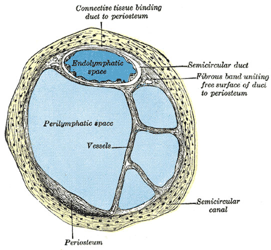
Human Semicircular Canal and Duct
Transverse section of a human semicircular canal and duct (after Rüdinger).
Structure
The walls of the utricle, saccule, and semicircular ducts consist of three layers.
- The outer layer is a loose and flocculent structure, apparently composed of ordinary fibrous tissue containing bloodvessels and some pigment-cells.
- The middle layer, thicker and more transparent, forms a homogeneous membrana propria, and presents on its internal surface, especially in the semicircular ducts, numerous papilliform projections, which, on the addition of acetic acid, exhibit an appearance of longitudinal fibrillation.
- The inner layer is formed of polygonal nucleated epithelial cells.
In the maculæ of the utricle and saccule, and in the transverse septa of the ampullæ of the semicircular ducts, the middle coat is thickened and the epithelium is columnar, and consists of supporting cells and hair cells. The former are fusiform, and their deep ends are attached to the membrana propria, while their free extremities are united to form a thin cuticle.
The hair cells are flask-shaped, and their deep, rounded ends do not reach the membrana propria, but lie between the supporting cells. The deep part of each contains a large nucleus, while its more superficial part is granular and pigmented. The free end is surmounted by a long, tapering, hair-like filament, which projects into the cavity. The filaments of the acoustic nerve enter these parts, and having pierced the outer and middle layers, they lose their medullary sheaths, and their axis-cylinders ramify between the hair cells.
(Text modified from Gray's 1918 Anatomy)
- Gray's Images: Development | Lymphatic | Neural | Vision | Hearing | Somatosensory | Integumentary | Respiratory | Gastrointestinal | Urogenital | Endocrine | Surface Anatomy | iBook | Historic Disclaimer
| Historic Disclaimer - information about historic embryology pages |
|---|
| Pages where the terms "Historic" (textbooks, papers, people, recommendations) appear on this site, and sections within pages where this disclaimer appears, indicate that the content and scientific understanding are specific to the time of publication. This means that while some scientific descriptions are still accurate, the terminology and interpretation of the developmental mechanisms reflect the understanding at the time of original publication and those of the preceding periods, these terms, interpretations and recommendations may not reflect our current scientific understanding. (More? Embryology History | Historic Embryology Papers) |
| iBook - Gray's Embryology | |
|---|---|

|
|
Reference
Gray H. Anatomy of the human body. (1918) Philadelphia: Lea & Febiger.
Cite this page: Hill, M.A. (2024, April 26) Embryology Gray0927.jpg. Retrieved from https://embryology.med.unsw.edu.au/embryology/index.php/File:Gray0927.jpg
- © Dr Mark Hill 2024, UNSW Embryology ISBN: 978 0 7334 2609 4 - UNSW CRICOS Provider Code No. 00098G
File history
Click on a date/time to view the file as it appeared at that time.
| Date/Time | Thumbnail | Dimensions | User | Comment | |
|---|---|---|---|---|---|
| current | 07:46, 19 August 2012 |  | 542 × 500 (82 KB) | Z8600021 (talk | contribs) | Transverse section of a human semicircular canal and duct (after Rüdinger). |
You cannot overwrite this file.
File usage
The following page uses this file:

