File:Gray0926.jpg
Gray0926.jpg (719 × 500 pixels, file size: 90 KB, MIME type: image/jpeg)
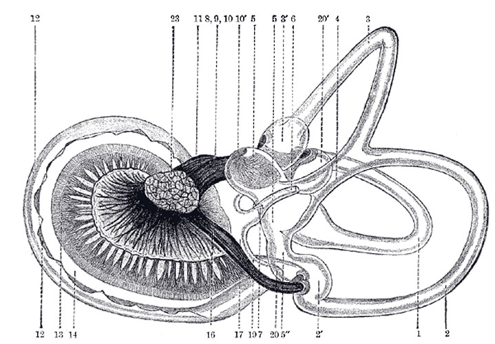
Right Human Membranous Labyrinth
The same as Fig. 925 from the postero-medial aspect. (G. Retzius.)
Legend
|
1. Lateral semicircular canal 1’, its ampulla 2. Posterior canal 2’, its ampulla. 3. Superior canal; 3’, its ampulla. 4. Conjoined limb of superior and posterior canals (sinus utriculi superior). 5. Utricle. 5’. Recessus utriculi. 5”. Sinus utriculi posterior. 6. Ductus endolymphaticus. 7. Canalis utriculosaccularis. 8. Nerve to ampulla of superior canal. 9. Nerve to ampulla of lateral canal. 10. Nerve to recessus utriculi (in Fig. 925, the three branches appear conjoined). 10’. Ending of nerve in recessus utriculi. |
11. Facial nerve. 12. Lagena cochleæ. 13. Nerve of cochlea within spiral lamina. 14. Basilar membrane. 15. Nerve fibers to macula of saccule. 16. Nerve to ampulla of posterior canal. 17. Saccule. 18. Secondary membrane of tympanum. 19. Canalis reuniens. 20. Vestibular end of ductus cochlearis. 23. Section of the facial and acoustic nerves within internal acoustic meatus (the separation between them is not apparent in the section). |
The Semicircular Ducts
(ductus semicirculares; membranous semicircular canals), (Fig. 925, Fig. 926).—The semicircular ducts are about one-fourth of the diameter of the osseous canals, but in number, shape, and general form they are precisely similar, and each presents at one end an ampulla. They open by five orifices into the utricle, one opening being common to the medial end of the superior and the upper end of the posterior duct. In the ampullæ the wall is thickened, and projects into the cavity as a fiddle-shaped, transversely placed elevation, the septum transversum, in which the nerves end.
The utricle, saccule, and semicircular ducts are held in position by numerous fibrous bands which stretch across the space between them and the bony walls.
(Text modified from Gray's 1918 Anatomy)
- Gray's Images: Development | Lymphatic | Neural | Vision | Hearing | Somatosensory | Integumentary | Respiratory | Gastrointestinal | Urogenital | Endocrine | Surface Anatomy | iBook | Historic Disclaimer
| Historic Disclaimer - information about historic embryology pages |
|---|
| Pages where the terms "Historic" (textbooks, papers, people, recommendations) appear on this site, and sections within pages where this disclaimer appears, indicate that the content and scientific understanding are specific to the time of publication. This means that while some scientific descriptions are still accurate, the terminology and interpretation of the developmental mechanisms reflect the understanding at the time of original publication and those of the preceding periods, these terms, interpretations and recommendations may not reflect our current scientific understanding. (More? Embryology History | Historic Embryology Papers) |
| iBook - Gray's Embryology | |
|---|---|

|
|
Reference
Gray H. Anatomy of the human body. (1918) Philadelphia: Lea & Febiger.
Cite this page: Hill, M.A. (2024, April 26) Embryology Gray0926.jpg. Retrieved from https://embryology.med.unsw.edu.au/embryology/index.php/File:Gray0926.jpg
- © Dr Mark Hill 2024, UNSW Embryology ISBN: 978 0 7334 2609 4 - UNSW CRICOS Provider Code No. 00098G
File history
Click on a date/time to view the file as it appeared at that time.
| Date/Time | Thumbnail | Dimensions | User | Comment | |
|---|---|---|---|---|---|
| current | 07:44, 19 August 2012 |  | 719 × 500 (90 KB) | Z8600021 (talk | contribs) | The same from the postero-medial aspect. 1. Lateral semicircular canal; 1’, its ampulla; 2. Posterior canal; 2’, its ampulla. 3. Superior canal; 3’, its ampulla. 4. Conjoined limb of superior and posterior canals (sinus utriculi superior). 5. Utricl |
You cannot overwrite this file.
File usage
The following page uses this file:

