File:Gray0923.jpg
Gray0923.jpg (691 × 500 pixels, file size: 106 KB, MIME type: image/jpeg)
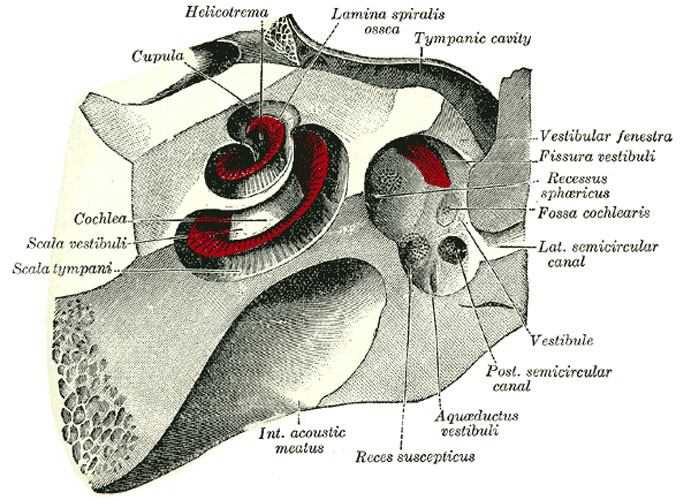
Inner Ear - cochlea and vestibule
The cochlea and vestibule, viewed from above. All the hard parts which form the roof of the internal ear have been removed with the saw.
The Cochlea (Figs. 922, 923).—The cochlea bears some resemblance to a common snail-shell; it forms the anterior part of the labyrinth, is conical in form, and placed almost horizontally in front of the vestibule; its apex (cupula) is directed forward and lateralward, with a slight inclination downward, toward the upper and front part of the labyrinthic wall of the tympanic cavity; its base corresponds with the bottom of the internal acoustic meatus, and is perforated by numerous apertures for the passage of the cochlear division of the acoustic nerve. It measures about 5 mm. from base to apex, and its breadth across the base is about 9 mm. It consists of a conical shaped central axis, the modiolus; of a canal, the inner wall of which is formed by the central axis, wound spirally around it for two turns and three-quarters, from the base to the apex; and of a delicate lamina, the osseous spiral lamina, which projects from the modiolus, and, following the windings of the canal, partially subdivides it into two. In the recent state a membrane, the basilar membrane, stretches from the free border of this lamina to the outer wall of the bony cochlea and completely separates the canal into two passages, which, however, communicate with each other at the apex of the modiolus by a small opening named the helicotrema.
The modiolus is the conical central axis or pillar of the cochlea. Its base is broad, and appears at the bottom of the internal acoustic meatus, where it corresponds with the area cochleæ; it is perforated by numerous orifices, which transmit filaments of the cochlear division of the acoustic nerve; the nerves for the first turn and a half pass through the foramina of the tractus spiralis foraminosus; those for the apical turn, through the foramen centrale. The canals of the tractus spiralis foraminosus pass up through the modiolus and successively bend outward to reach the attached margin of the lamina spiralis ossea. Here they become enlarged, and by their apposition form the spiral canal of the modiolus, which follows the course of the attached margin of the osseous spiral lamina and lodges the spiral ganglion (ganglion of Corti). The foramen centrale is continued into a canal which runs up the middle of the modiolus to its apex. The modiolus diminishes rapidly in size in the second and succeeding coil.
The bony canal of the cochlea takes two turns and three-quarters around the modiolus. It is about 30 mm. in length, and diminishes gradually in diameter from the base to the summit, where it terminates in the cupula, which forms the apex of the cochlea. The beginning of this canal is about 3 mm. in diameter; it diverges from the modiolus toward the tympanic cavity and vestibule, and presents three openings. One, the fenestra cochleæ, communicates with the tympanic cavity—in the fresh state this aperture is closed by the secondary tympanic membrane; another, of an elliptical form, opens into the vestibule. The third is the aperture of the aquæductus cochleæ, leading to a minute funnel-shaped canal, which opens on the inferior surface of the petrous part of the temporal bone and transmits a small vein, and also forms a communication between the subarachnoid cavity and the scala tympani.
(Text modified from Gray's 1918 Anatomy)
- Gray's Images: Development | Lymphatic | Neural | Vision | Hearing | Somatosensory | Integumentary | Respiratory | Gastrointestinal | Urogenital | Endocrine | Surface Anatomy | iBook | Historic Disclaimer
| Historic Disclaimer - information about historic embryology pages |
|---|
| Pages where the terms "Historic" (textbooks, papers, people, recommendations) appear on this site, and sections within pages where this disclaimer appears, indicate that the content and scientific understanding are specific to the time of publication. This means that while some scientific descriptions are still accurate, the terminology and interpretation of the developmental mechanisms reflect the understanding at the time of original publication and those of the preceding periods, these terms, interpretations and recommendations may not reflect our current scientific understanding. (More? Embryology History | Historic Embryology Papers) |
| iBook - Gray's Embryology | |
|---|---|

|
|
Reference
Gray H. Anatomy of the human body. (1918) Philadelphia: Lea & Febiger.
Cite this page: Hill, M.A. (2024, April 26) Embryology Gray0923.jpg. Retrieved from https://embryology.med.unsw.edu.au/embryology/index.php/File:Gray0923.jpg
- © Dr Mark Hill 2024, UNSW Embryology ISBN: 978 0 7334 2609 4 - UNSW CRICOS Provider Code No. 00098G
File history
Click on a date/time to view the file as it appeared at that time.
| Date/Time | Thumbnail | Dimensions | User | Comment | |
|---|---|---|---|---|---|
| current | 05:08, 19 August 2012 |  | 691 × 500 (106 KB) | Z8600021 (talk | contribs) | ==Inner Ear - cochlea and vestibule== The cochlea and vestibule, viewed from above. All the hard parts which form the roof of the internal ear have been removed with the saw. The Cochlea (Figs. 922, 923).—The cochlea bears some resemblance to a common |
You cannot overwrite this file.
File usage
The following page uses this file:

