File:Gray0921.jpg
Gray0921.jpg (640 × 500 pixels, file size: 95 KB, MIME type: image/jpeg)
Osseous Labyrinth
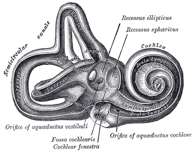
Right osseous labyrinth, Interior.
The Osseous Labyrinth (labyrinthus osseus) (Figs. 920, 921).—The osseous labyrinth consists of three parts: the vestibule, semicircular canals, and cochlea. These are cavities hollowed out of the substance of the bone, and lined by periosteum; they contain a clear fluid, the perilymph, in which the membranous labyrinth is situated.
The Vestibule (vestibulum).—The vestibule is the central part of the osseous labyrinth, and is situated medial to the tympanic cavity, behind the cochlea, and in front of the semicircular canals. It is somewhat ovoid in shape, but flattened transversely; it measures about 5 mm. from before backward, the same from above downward, and about 3 mm. across. In its lateral or tympanic wall is the fenestra vestibuli, closed, in the fresh state, by the base of the stapes and annular ligament. On its medial wall, at the forepart, is a small circular depression, the recessus sphæricus, which is perforated, at its anterior and inferior part, by several minute holes (macula cribrosa media) for the passage of filaments of the acoustic nerve to the saccule; and behind this depression is an oblique ridge, the crista vestibuli, the anterior end of which is named the pyramid of the vestibule. This ridge bifurcates below to enclose a small depression, the fossa cochlearis, which is perforated by a number of holes for the passage of filaments of the acoustic nerve which supply the vestibular end of the ductus cochlearis. As the hinder part of the medial wall is the orifice of the aquæductus vestibuli, which extends to the posterior surface of the petrous portion of the temporal bone. It transmits a small vein, and contains a tubular prolongation of the membranous labyrinth, the ductus endolymphaticus, which ends in a cul-de-sac between the layers of the dura mater within the cranial cavity. On the upper wall or roof is a transversely oval depression, the recessus ellipticus, separated from the recessus sphæricus by the crista vestibuli already mentioned. The pyramid and adjoining part of the recessus ellipticus are perforated by a number of holes (macula cribrosa superior). The apertures in the pyramid transmit the nerves to the utricle; those in the recessus ellipticus the nerves to the ampullæ of the superior and lateral semicircular ducts. Behind are the five orifices of the semicircular canals. In front is an elliptical opening, which communicates with the scala vestibuli of the cochlea.
(Text modified from Gray's 1918 Anatomy)
- Gray's Images: Development | Lymphatic | Neural | Vision | Hearing | Somatosensory | Integumentary | Respiratory | Gastrointestinal | Urogenital | Endocrine | Surface Anatomy | iBook | Historic Disclaimer
| Historic Disclaimer - information about historic embryology pages |
|---|
| Pages where the terms "Historic" (textbooks, papers, people, recommendations) appear on this site, and sections within pages where this disclaimer appears, indicate that the content and scientific understanding are specific to the time of publication. This means that while some scientific descriptions are still accurate, the terminology and interpretation of the developmental mechanisms reflect the understanding at the time of original publication and those of the preceding periods, these terms, interpretations and recommendations may not reflect our current scientific understanding. (More? Embryology History | Historic Embryology Papers) |
| iBook - Gray's Embryology | |
|---|---|

|
|
Reference
Gray H. Anatomy of the human body. (1918) Philadelphia: Lea & Febiger.
Cite this page: Hill, M.A. (2024, April 24) Embryology Gray0921.jpg. Retrieved from https://embryology.med.unsw.edu.au/embryology/index.php/File:Gray0921.jpg
- © Dr Mark Hill 2024, UNSW Embryology ISBN: 978 0 7334 2609 4 - UNSW CRICOS Provider Code No. 00098G
File history
Click on a date/time to view the file as it appeared at that time.
| Date/Time | Thumbnail | Dimensions | User | Comment | |
|---|---|---|---|---|---|
| current | 07:26, 19 August 2012 |  | 640 × 500 (95 KB) | Z8600021 (talk | contribs) |
You cannot overwrite this file.
File usage
The following page uses this file:

