File:Gray0892.jpg
Gray0892.jpg (500 × 409 pixels, file size: 47 KB, MIME type: image/jpeg)
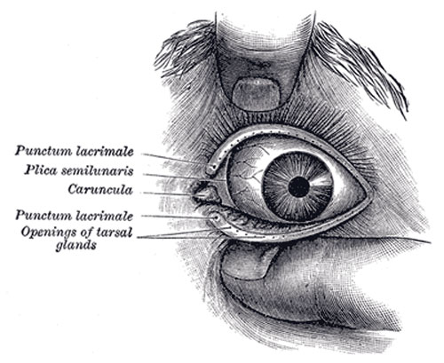
Front of left eye with eyelids separated to show medial canthus
The Eyelids (palpebræ) are two thin, movable folds, placed in front of the eye, protecting it from injury by their closure. The upper eyelid is the larger, and the more movable of the two, and is furnished with an elevator muscle, the Levator palpebræ superioris. When the eyelids are open, an elliptical space, the palpebral fissure (rima palpebrarum), is left between their margins, the angles of which correspond to the junctions of the upper and lower eyelids, and are called the palpebral commissures or canthi.
The lateral palpebral commissure (commissura palpebrarum lateralis; external canthus) is more acute than the medial, and the eyelids here lie in close contact with the bulb of the eye: but the medial palpebral commissure (commissura palpebrarum medialis; internal canthus) is prolonged for a short distance toward the nose, and the two eyelids are separated by a triangular space, the lacus lacrimalis (Fig. 892). At the basal angles of the lacus lacrimalis, on the margin of each eyelid, is a small conical elevation, the lacrimal papilla, the apex of which is pierced by a small orifice, the punctum lacrimale, the commencement of the lacrimal duct.
The eyelashes (cilia) are attached to the free edges of the eyelids; they are short, thick, curved hairs, arranged in a double or triple row: those of the upper eyelid, more numerous and longer than those of the lower, curve upward; those of the lower eyelid curve downward, so that they do not interlace in closing the lids. Near the attachment of the eyelashes are the openings of a number of glands, the ciliary glands, arranged in several rows close to the free margin of the lid; they are regarded as enlarged and modified sudoriferous glands.
(Text modified from Gray's 1918 Anatomy)
- Gray's Images: Development | Lymphatic | Neural | Vision | Hearing | Somatosensory | Integumentary | Respiratory | Gastrointestinal | Urogenital | Endocrine | Surface Anatomy | iBook | Historic Disclaimer
| Historic Disclaimer - information about historic embryology pages |
|---|
 |
| iBook - Gray's Embryology | |
|---|---|

|
|
Reference
Gray H. Anatomy of the human body. (1918) Philadelphia: Lea & Febiger.
Cite this page: Hill, M.A. (2024, April 26) Embryology Gray0892.jpg. Retrieved from https://embryology.med.unsw.edu.au/embryology/index.php/File:Gray0892.jpg
- © Dr Mark Hill 2024, UNSW Embryology ISBN: 978 0 7334 2609 4 - UNSW CRICOS Provider Code No. 00098G
File history
Click on a date/time to view the file as it appeared at that time.
| Date/Time | Thumbnail | Dimensions | User | Comment | |
|---|---|---|---|---|---|
| current | 22:49, 19 August 2012 | | 500 × 409 (47 KB) | Z8600021 (talk | contribs) | |
| 09:53, 9 March 2011 |  | 400 × 327 (36 KB) | S8600021 (talk | contribs) | {{Gray Anatomy}} |
You cannot overwrite this file.
File usage
The following 2 pages use this file: