File:Gray0855.jpg
Gray0855.jpg (500 × 468 pixels, file size: 108 KB, MIME type: image/jpeg)
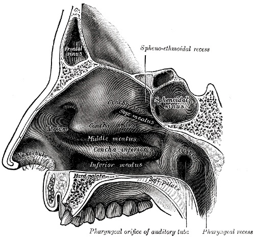
Fig. 855. Lateral Wall of Nasal Cavity
Palate Development | Respiratory Development | Smell Development
Lateral Wall (Figs. 855, 856). On the lateral wall are the superior, middle, and inferior nasal conchæ, and below and lateral to each concha is the corresponding nasal passage or meatus. Above the superior concha is a narrow recess, the sphenoethmoidal recess, into which the sphenoidal sinus opens. The superior meatus is a short oblique passage extending about half-way along the upper border of the middle concha; the posterior ethmoidal cells open into the front part of this meatus. The middle meatus is below and lateral to the middle concha, and is continued anteriorly into a shallow depression, situated above the vestibule and named the atrium of the middle meatus. On raising or removing the middle concha the lateral wall of this meatus is fully displayed. On it is a rounded elevation, the bulla ethmoidalis, and below and in front of this is a curved cleft, the hiatus semilunaris.
The floor is concave from side to side and almost horizontal antero-posteriorly; its anterior three-fourths are formed by the palatine process of the maxilla, its posterior fourth by the horizontal process of the palatine bone. In its anteromedial part, directly over the incisive foramen, a small depression, the nasopalatine recess, is sometimes seen; it points downward and forward and occupies the position of a canal which connected the nasal with the buccal cavity in early fetal life.
- Gray's Images: Development | Lymphatic | Neural | Vision | Hearing | Somatosensory | Integumentary | Respiratory | Gastrointestinal | Urogenital | Endocrine | Surface Anatomy | iBook | Historic Disclaimer
| Historic Disclaimer - information about historic embryology pages |
|---|
| Pages where the terms "Historic" (textbooks, papers, people, recommendations) appear on this site, and sections within pages where this disclaimer appears, indicate that the content and scientific understanding are specific to the time of publication. This means that while some scientific descriptions are still accurate, the terminology and interpretation of the developmental mechanisms reflect the understanding at the time of original publication and those of the preceding periods, these terms, interpretations and recommendations may not reflect our current scientific understanding. (More? Embryology History | Historic Embryology Papers) |
| iBook - Gray's Embryology | |
|---|---|

|
|
Reference
Gray H. Anatomy of the human body. (1918) Philadelphia: Lea & Febiger.
Cite this page: Hill, M.A. (2024, April 26) Embryology Gray0855.jpg. Retrieved from https://embryology.med.unsw.edu.au/embryology/index.php/File:Gray0855.jpg
- © Dr Mark Hill 2024, UNSW Embryology ISBN: 978 0 7334 2609 4 - UNSW CRICOS Provider Code No. 00098G
File history
Click on a date/time to view the file as it appeared at that time.
| Date/Time | Thumbnail | Dimensions | User | Comment | |
|---|---|---|---|---|---|
| current | 22:19, 13 May 2013 |  | 500 × 468 (108 KB) | Z8600021 (talk | contribs) | {{Gray Anatomy}} |
You cannot overwrite this file.
File usage
The following 2 pages use this file:

