File:Gray0769.jpg
Gray0769.jpg (600 × 454 pixels, file size: 101 KB, MIME type: image/jpeg)
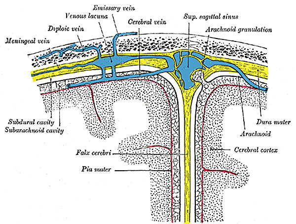
Fig. 769. Diagrammatic representation of a section across the top of the skull
Showing the membranes of the brain, etc. (Modified from Testut.)
The Arachnoid Villi (granulationes arachnoideales; glandulæ Pacchioni; Pacchionian bodies) (Fig. 769) are small, fleshy-looking elevations, usually collected into clusters of variable size, which are present upon the outer surface of the dura mater, in the vicinity of the superior sagittal sinus, and in some other situations. Upon laying open the sagittal sinus and the venous lacunæ on either side of it villi will be found protruding into its interior. They are not seen in infancy, and very rarely until the third year. They are usually found after the seventh year; and from this period they increase in number and size as age advances. They are not glandular in structure, but are enlarged normal villi of the arachnoid. As they grow they push the thinned dura mater before them, and cause absorption of the bone from pressure, and so produce the pits or depressions on the inner wall of the calvarium.
(text modified from Gray's Anatomy)
- Gray's Images: Development | Lymphatic | Neural | Vision | Hearing | Somatosensory | Integumentary | Respiratory | Gastrointestinal | Urogenital | Endocrine | Surface Anatomy | iBook | Historic Disclaimer
| Historic Disclaimer - information about historic embryology pages |
|---|
 |
| iBook - Gray's Embryology | |
|---|---|

|
|
Reference
Gray H. Anatomy of the human body. (1918) Philadelphia: Lea & Febiger.
Cite this page: Hill, M.A. (2024, April 26) Embryology Gray0769.jpg. Retrieved from https://embryology.med.unsw.edu.au/embryology/index.php/File:Gray0769.jpg
- © Dr Mark Hill 2024, UNSW Embryology ISBN: 978 0 7334 2609 4 - UNSW CRICOS Provider Code No. 00098G
File history
Click on a date/time to view the file as it appeared at that time.
| Date/Time | Thumbnail | Dimensions | User | Comment | |
|---|---|---|---|---|---|
| current | 10:12, 6 February 2016 | | 600 × 454 (101 KB) | Z8600021 (talk | contribs) |
You cannot overwrite this file.
File usage
The following 2 pages use this file: