File:Gray0715.jpg
Gray0715.jpg (800 × 591 pixels, file size: 127 KB, MIME type: image/jpeg)
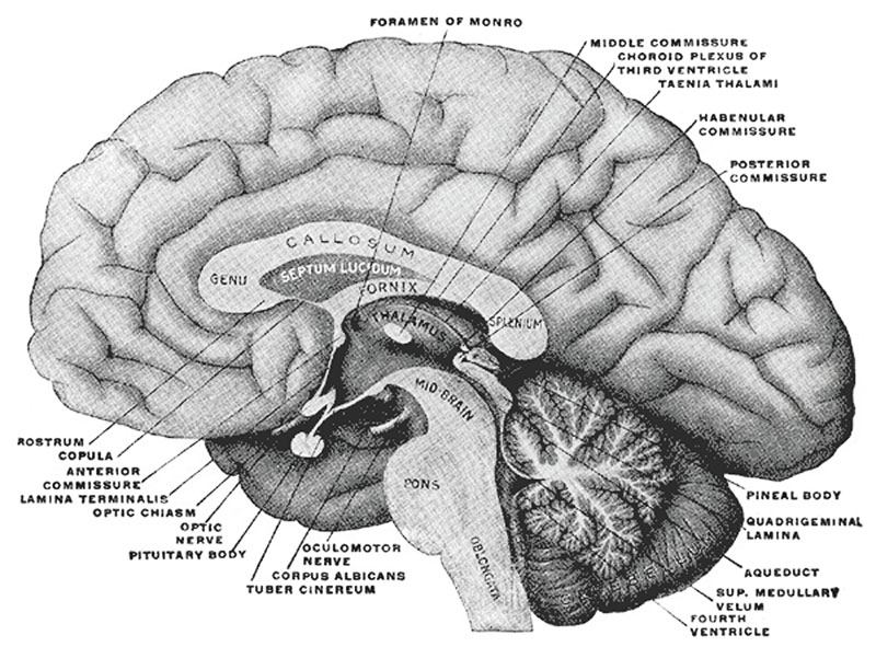
Fig. 715. Mesial aspect of a brain sectioned in the median sagittal plan
The Diencephalon
The diencephalon is connected above and in front with the cerebral hemispheres; behind with the mid-brain. Its upper surface is concealed by the corpus callosum, and is covered by a fold of pia mater, named the tela chorioidea of the third ventricle; inferiorly it reaches to the base of the brain.
The diencephalon comprises:
- thalamencephalon
- pars mamillaris hypothalami
- posterior part of the third ventricle.
For descriptive purposes, however, it is more convenient to consider the whole of the third ventricle and its boundaries together; this necessitates the inclusion, under this heading, of the pars optica hypothalami and the corresponding part of the third ventricle—structures which properly belong to the telencephalon.
- Gray's Images: Development | Lymphatic | Neural | Vision | Hearing | Somatosensory | Integumentary | Respiratory | Gastrointestinal | Urogenital | Endocrine | Surface Anatomy | iBook | Historic Disclaimer
| Historic Disclaimer - information about historic embryology pages |
|---|
 |
| iBook - Gray's Embryology | |
|---|---|

|
|
Reference
Gray H. Anatomy of the human body. (1918) Philadelphia: Lea & Febiger.
Cite this page: Hill, M.A. (2024, April 27) Embryology Gray0715.jpg. Retrieved from https://embryology.med.unsw.edu.au/embryology/index.php/File:Gray0715.jpg
- © Dr Mark Hill 2024, UNSW Embryology ISBN: 978 0 7334 2609 4 - UNSW CRICOS Provider Code No. 00098G
File history
Click on a date/time to view the file as it appeared at that time.
| Date/Time | Thumbnail | Dimensions | User | Comment | |
|---|---|---|---|---|---|
| current | 15:37, 15 May 2012 | | 800 × 591 (127 KB) | Z8600021 (talk | contribs) |
You cannot overwrite this file.
File usage
The following page uses this file: