File:Frazer1928 fig06.jpg

Original file (1,000 × 474 pixels, file size: 104 KB, MIME type: image/jpeg)
Text-fig. 6. Schematic analysis 35 mm and 48 mm
35 mm on left. Sections A 35 mm., B 48 mm
Text-fig. 6 gives the schematic analysis of the 35 mm. stage, with a halfsection (A). It can be seen that the basal wall of the cavity is practically as before, save that there is now no sign of the intralaminar remnant, the trans-laminar being represented alone as a deep groove. The lower part of the basal area has thickened considerably, with migration of neuroblasts. The main feature, however, is the great increase in size of the alar area, which now extends down well over the outer part of the basal formation.
The second half-section, B, represents the conditions in the 48 mm. specimen, at a much smaller magnification. The alar growth is now larger and a ventro-lateral extension of it is shown: whether this is really alar in formation, or a secondary involvement of the basal surface by fibres running to the alar structures, cannot be decided, although the latter seems to me to be the more likely explanation. Fibres extending (superior peduncle) into the basal region from the alar, are clearly seen: these, as a matter of fact, are also suggested to some extent in the 35 mm. embryo. The ventro-lateral fibres seem to belong to the lateral lemniscus formation.
The position of the groups or cells is now shown in the section (text—fig. 6) by large black dots. This clearly represents the mesencephalic root of the nerve, and it extends forward at this stage only as far as the alar invasion seems to have extended, that is, to the deep hollow in front of the invaginated parts.
| Historic Disclaimer - information about historic embryology pages |
|---|
 |
Reference
Frazer JE. Development of the region of the isthmus rhombencephali. (1928) J Anat. 63: 7-18. PMID 17104212
Cite this page: Hill, M.A. (2024, April 26) Embryology Frazer1928 fig06.jpg. Retrieved from https://embryology.med.unsw.edu.au/embryology/index.php/File:Frazer1928_fig06.jpg
- © Dr Mark Hill 2024, UNSW Embryology ISBN: 978 0 7334 2609 4 - UNSW CRICOS Provider Code No. 00098G
File history
Click on a date/time to view the file as it appeared at that time.
| Date/Time | Thumbnail | Dimensions | User | Comment | |
|---|---|---|---|---|---|
| current | 13:16, 9 January 2017 | | 1,000 × 474 (104 KB) | Z8600021 (talk | contribs) | |
| 13:15, 9 January 2017 | 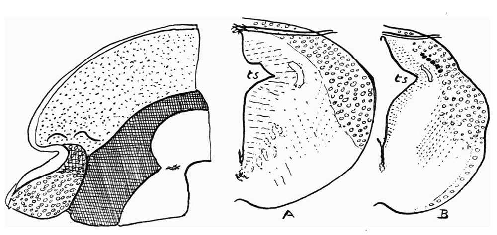 | 1,000 × 474 (104 KB) | Z8600021 (talk | contribs) | ||
| 13:11, 9 January 2017 | 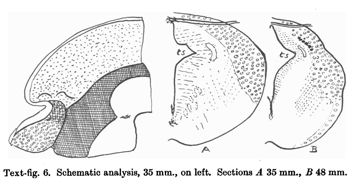 | 1,214 × 624 (130 KB) | Z8600021 (talk | contribs) | {{Historic Disclaimer}} ===Reference=== {{Ref-Frazer1928}} {{Footer}} |
You cannot overwrite this file.
File usage
The following page uses this file: