File:Frazer1910 fig08.jpg

Original file (1,300 × 708 pixels, file size: 202 KB, MIME type: image/jpeg)
Fig. 8. Reconstruction model embryo 35 mm
A is a reduced drawing of the model from behind.
B is a larger drawing of the model from the left and below, the lateral piece being removed with the thyroid ala. The chordal nodule (Ch. N.) is seen connected below with the cricoid, which is very large. The saccular outgrowth (S¢wc.) has the false cord above it, and, above this, the lateral edge of the wall of the transverse cavity is seen. Part of the wall of the sagittal cavity is shown (s) behind the arytaenoid, and a little of this wall is exposed behind and below the chordal nodule, where the original connection of the nodule with the cricoid has thinned out, and is no longer demonstrable, leaving the wall exposed.
C. A drawing of a separate model of the cavity oi the same embryo, divided mesially and seen from the right.
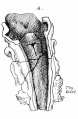
8 A model from behind
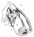
8 B model from the left and below
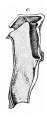
8 C model of the cavity


Online Editor - Embryo CRL 35 mm can be Week 9 early fetal period.
| Historic Disclaimer - information about historic embryology pages |
|---|
 |
- Links: fig 1 | fig 2 | fig 3 | fig 4 | fig 5 | fig 6 | fig 7 | fig 8 | fig 9 | fig 10 | fig 11 | fig 12 | fig 13 | fig 14 | fig 15 | fig 16 | fig 17 | fig 18 | fig 19 | 1910 Frazer | Historic Embryology Papers | Respiratory System Development
Reference
Frazer JE. Development of the larynx. (1910) J Anat. 44: 156-191. PMID 17232839
Cite this page: Hill, M.A. (2024, April 26) Embryology Frazer1910 fig08.jpg. Retrieved from https://embryology.med.unsw.edu.au/embryology/index.php/File:Frazer1910_fig08.jpg
- © Dr Mark Hill 2024, UNSW Embryology ISBN: 978 0 7334 2609 4 - UNSW CRICOS Provider Code No. 00098G
File history
Click on a date/time to view the file as it appeared at that time.
| Date/Time | Thumbnail | Dimensions | User | Comment | |
|---|---|---|---|---|---|
| current | 09:20, 11 January 2017 | | 1,300 × 708 (202 KB) | Z8600021 (talk | contribs) | |
| 09:14, 11 January 2017 |  | 1,567 × 1,220 (376 KB) | Z8600021 (talk | contribs) | {{Frazer1910 figures}} |
You cannot overwrite this file.
File usage
The following 4 pages use this file: