File:Fetal kidney MRI 02.jpg

Original file (797 × 880 pixels, file size: 64 KB, MIME type: image/jpeg)
Fetal Kidney Magnetic Resonance Image
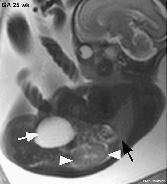
MRI appearance of normal fetal kidneys. Sagittal T2- SSFSE of a fetal abdomen at 25 WG: Note the size and the signal appearance of the normal kidney between the two arrowheads. The fluid-filled urinary bladder (white arrow), the adequate volume of the amniotic fluid, and the developing lungs (black arrow) indicate good renal function. Note that the urinary bladder can occupy a considerable portion of the abdomen as a normal finding.
- MRI Links: image - renal labeled 25wk | image - renal 25wk | Renal Development | Magnetic Resonance Imaging
Reference
<pubmed>25685519</pubmed>| J Adv Res.
Copyright
Image source: The MRI images are reproduced with the permission of Prof Sahar Saleem for educational purposes only and cannot be reproduced electronically or in writing without permission. http://creativecommons.org/licenses/by-nc-nd/3.0/
Fig. 19 Gr19.jpg 1-s2.0-S2090123213000805-g192.jpg Original image reorganised in size and relabeled.
File history
Click on a date/time to view the file as it appeared at that time.
| Date/Time | Thumbnail | Dimensions | User | Comment | |
|---|---|---|---|---|---|
| current | 15:11, 1 March 2015 |  | 797 × 880 (64 KB) | Z8600021 (talk | contribs) | ==Fetal Kidney Magnetic Resonance Image== MRI appearance of normal fetal kidneys. Sagittal T2- SSFSE of a fetal abdomen at 25 WG: Note the size and the signal appearance of the normal kidney between the two arrowheads. The fluid-filled urinary bladder... |
You cannot overwrite this file.
File usage
There are no pages that use this file.