File:Bovine blastocyst KRT18, FN1 and MYL6 expression.jpg

Original file (1,000 × 499 pixels, file size: 90 KB, MIME type: image/jpeg)
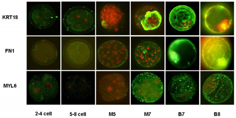
Bovine blastocyst (in vitro) KRT18, FN1 and MYL6 expression
Confocal laser scanning images of in vitro produced bovine embryos labelled with primary mouse antibodies for KRT18, FN1 and MYL6 respectively in combination with FITC-labelled secondary goat-anti-mouse antibodies.
The nuclei are stained with propidium iodide.
Different stages of preimplantation embryo development were analysed (2–4 cell, 5–8 cell, M5: morula day 5 p.i., M7: morula day 7 p.i., B7: blastocyst day 7 p.i., B8: blastocyst day 8 p.i.).
Original file name: Figure 2. 1471-213X-7-64-3.jpg http://www.biomedcentral.com/1471-213X/7/64/figure/F2
Reference
<pubmed>17559642</pubmed>| BMC Dev Biol.
Goossens et al. BMC Developmental Biology 2007 7:64 doi:10.1186/1471-213X-7-64
© 2007 Goossens et al; licensee BioMed Central Ltd. This is an Open Access article distributed under the terms of the Creative Commons Attribution License (http://creativecommons.org/licenses/by/2.0), which permits unrestricted use, distribution, and reproduction in any medium, provided the original work is properly cited.
File history
Click on a date/time to view the file as it appeared at that time.
| Date/Time | Thumbnail | Dimensions | User | Comment | |
|---|---|---|---|---|---|
| current | 15:29, 14 October 2010 | | 1,000 × 499 (90 KB) | S8600021 (talk | contribs) | ==Bovine blastocyst (in vitro) KRT18, FN1 and MYL6 expression== Confocal laser scanning images of in vitro produced bovine embryos labelled with primary mouse antibodies for KRT18, FN1 and MYL6 respectively in combination with FITC-labelled secondary go |
You cannot overwrite this file.
File usage
The following page uses this file: