File:Baboon- fetal brain.jpg

Original file (1,000 × 733 pixels, file size: 127 KB, MIME type: image/jpeg)
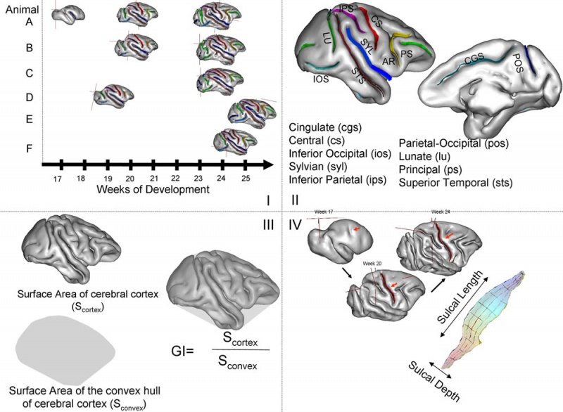
(I) Cortical surfaces for six fetuses (A–F).
(II) Ten primary sulci used for the analysis of the sulcal length and depth.
(III) Measurement of gyrification index (GI).
(IV) Measurements of sulcal length and depth.
Original File Name: Figure 2. Fnins-04-00020-g002.jpg http://www.frontiersin.org/TempImages/imagecache/1250_fnins-04-00020/images/image_m/fnins-04-00020-g002.jpg
Reference
<pubmed>20631812</pubmed>| Front Neurosci.
Copyright: © 2010 Kochunov, Castro, Davis, Dudley, Brewer, Zhang, Kroenke, Purdy, Fox, Simerly and Schatten. This is an open-access article subject to an exclusive license agreement between the authors and the Frontiers Research Foundation, which permits unrestricted use, distribution, and reproduction in any medium, provided the original authors and source are credited.
File history
Click on a date/time to view the file as it appeared at that time.
| Date/Time | Thumbnail | Dimensions | User | Comment | |
|---|---|---|---|---|---|
| current | 14:47, 15 October 2010 |  | 1,000 × 733 (127 KB) | S8600021 (talk | contribs) | (I) Cortical surfaces for six fetuses (A–F). (II) Ten primary sulci used for the analysis of the sulcal length and depth. (III) Measurement of gyrification index (GI). (IV) Measurements of sulcal length and depth. Original File Name: Figure 2. F |
You cannot overwrite this file.
File usage
The following page uses this file: