File:Anson-1934 fig01-07.jpg

Original file (1,280 × 1,506 pixels, file size: 210 KB, MIME type: image/jpeg)
Figs. 1 to 7. Tracings of endolymphatic duct and adjacent area of membranous labyrinth from Human series
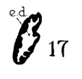
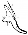
Figures 1 to 4 and 7, X 37; 5 and 6, X 25. 1, 22.8 mm.; 2, 29 mm.; 34, 40 mm.; 5, infant, 4 months; 6, child, 3 years; 7, 36 mm.
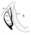
a.v., auditory vesicle; caps, cartilaginous otic capsule; c.d., cochlear duct; cochl., cochlear part of vesicle; div., saccular diverticula; e.ap., endolymphatic appendage; e.d., endolynlphatic duct; e.s., endolymphatie sac; ep., epidermis; l.s.d., lateral semicircular duct; m.t., medullary tube; sae., saccule; s.s.d., superior semicircular duct; st., stapes; utr., utricle; vest, vestibular part of vesicle; u.e.d., utriculo-endolymphatic duct; u.e.f., utriculur fold (‘valve); v., blood vessels. * Rugae, in tangential section. Numbered arrows designate folds discussed in text.
Online Editor Note the Harvard Collection human embryos were incorporated into the Carnegie Collection.
| Historic Disclaimer - information about historic embryology pages |
|---|
 |
- Links: fig 1-7 | fig 1 | fig 2 | fig 3 | fig 4 | fig 5 | fig 6 | fig 7 | fig 8-21 | fig 22-33 | 1934 Anson | Historic Papers | Inner Ear Development
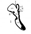
fig 1 Human 22.8 mm
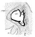
fig 2 Human 29 mm
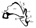
fig 3 Human 34 mm
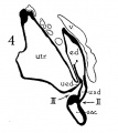
fig 4 Human 40 mm
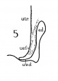
fig 5 Infant 4 months
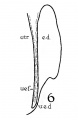
fig 6 Child 3 years
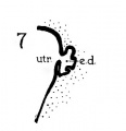
fig 7 embryo 36 mm
8 Cat 7 mm
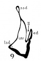
9 Cat 10.6 mm
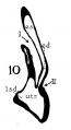
10 Cat 14 mm
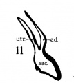
11 Cat 15 mm
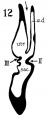
12 Cat 24.1 mm
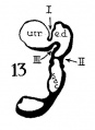
13 Cat 32.6 mm
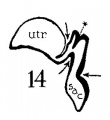
14 Cat 39 mm
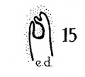
15 Cat 39 mm
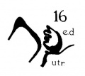
16 Cat 31 mm
17 Guinea-pig 18.5 mm
18 Dog 12.5 mm
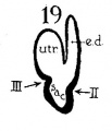
19 Rabbit 21 mm
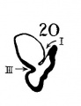
20 Rabbit 25 mm
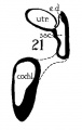
21 Rabbit 29 mm


Reference
Anson BJ. The early development of the membranous labyrinth in mammalian embryos, with special reference to the endolymphatic duct and the utriculo—endolymphatic duct. (1934) Anat. Rec. 59: 15-25.
Cite this page: Hill, M.A. (2024, April 26) Embryology Anson-1934 fig01-07.jpg. Retrieved from https://embryology.med.unsw.edu.au/embryology/index.php/File:Anson-1934_fig01-07.jpg
- © Dr Mark Hill 2024, UNSW Embryology ISBN: 978 0 7334 2609 4 - UNSW CRICOS Provider Code No. 00098G
File history
Click on a date/time to view the file as it appeared at that time.
| Date/Time | Thumbnail | Dimensions | User | Comment | |
|---|---|---|---|---|---|
| current | 09:36, 2 February 2017 | | 1,280 × 1,506 (210 KB) | Z8600021 (talk | contribs) | |
| 09:35, 2 February 2017 |  | 1,330 × 2,074 (378 KB) | Z8600021 (talk | contribs) | {{Anson1934 figures}} |
You cannot overwrite this file.
File usage
The following page uses this file: