Developmental Mechanism - Cell Migration

From Embryology
Embryology - 19 Mar 2024    Facebook link Pinterest link Twitter link  Expand to Translate  
Google Translate - select your language from the list shown below (this will open a new external page)

العربية | català | 中文 | 中國傳統的 | français | Deutsche | עִברִית | हिंदी | bahasa Indonesia | italiano | 日本語 | 한국어 | မြန်မာ | Pilipino | Polskie | português | ਪੰਜਾਬੀ ਦੇ | Română | русский | Español | Swahili | Svensk | ไทย | Türkçe | اردو | ייִדיש | Tiếng Việt    These external translations are automated and may not be accurate. (More? About Translations)

Introduction

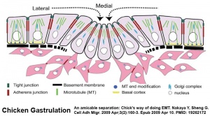
Gastrulation involves both epithelial to mesenchymal transition and cell migration

The process of cell migration occurs at different stages throughout embryonic development and involves other developmental mechanisms. The first key migration occurs during gastrulation. Later key migratory events also occur during somitogenesis and neural crest migration.

This mechanism involves several cellular processes including: cytoskeletal reorganisation, adhesion, extracellular matrix, and chemotactic signaling.


Mechanism - "a process, technique, or system for achieving a result"
Mechanism Links: mitosis | cell migration | cell junctions |epithelial invagination | epithelial mesenchymal transition | mesenchymal epithelial transition | epithelial mesenchymal interaction | morphodynamics | tube formation | apoptosis | autophagy | axes formation | time | molecular

Some Recent Findings

  • STRIP1, a core component of STRIPAK complexes, is essential for normal mesoderm migration in the mouse embryo[1] "Regulated mesoderm migration is necessary for the proper morphogenesis and organ formation during embryonic development. Cell migration and its dependence on the cytoskeleton and signaling machines have been studied extensively in cultured cells; in contrast, remarkably little is known about the mechanisms that regulate mesoderm cell migration in vivo. Here, we report the identification and characterization of a mouse mutation in striatin-interacting protein 1 (Strip1) that disrupts migration of the mesoderm after the gastrulation epithelial-to-mesenchymal transition (EMT). STRIP1 is a core component of the biochemically defined mammalian striatin-interacting phosphatases and kinase (STRIPAK) complexes that appear to act through regulation of protein phosphatase 2A (PP2A), but their functions in mammals in vivo have not been examined. Strip1-null mutants arrest development at midgestation with profound disruptions in the organization of the mesoderm and its derivatives, including a complete failure of the anterior extension of axial mesoderm. Analysis of cultured mesoderm explants and mouse embryonic fibroblasts from null mutants shows that the mesoderm migration defect is correlated with decreased cell spreading, abnormal focal adhesions, changes in the organization of the actin cytoskeleton, and decreased velocity of cell migration. The results show that STRIPAK complexes are essential for cell migration and tissue morphogenesis in vivo." (More? Mesoderm | NCBI Gene - STRIP1)
  • LKB1 signaling in cephalic neural crest cells is essential for vertebrate head development[2] "Head development in vertebrates proceeds through a series of elaborate patterning mechanisms and cell-cell interactions involving cephalic neural crest cells (CNCC). These cells undergo extensive migration along stereotypical paths after their separation from the dorsal margins of the neural tube and they give rise to most of the craniofacial skeleton. Here, we report that the silencing of the LKB1 tumor suppressor affects the delamination of pre-migratory CNCC from the neural primordium as well as their polarization and survival, thus resulting in severe facial and brain defects. We further show that LKB1-mediated effects on the development of CNCC involve the sequential activation of the AMP-activated protein kinase (AMPK), the Rho-dependent kinase (ROCK) and the actin-based motor protein myosin II. Collectively, these results establish that the complex morphogenetic processes governing head formation critically depends on the activation of the LKB1 signaling network in CNCC."
  • Review - Collective cell migration in development[3] "During embryonic development, tissues undergo major rearrangements that lead to germ layer positioning, patterning, and organ morphogenesis. Often these morphogenetic movements are accomplished by the coordinated and cooperative migration of the constituent cells, referred to as collective cell migration. The molecular and biomechanical mechanisms underlying collective migration of developing tissues have been investigated in a variety of models, including border cell migration, tracheal branching, blood vessel sprouting, and the migration of the lateral line primordium, neural crest cells, or head mesendoderm. Here we review recent advances in understanding collective migration in these developmental models, focusing on the interaction between cells and guidance cues presented by the microenvironment and on the role of cell-cell adhesion in mechanical and behavioral coupling of cells within the collective."
More recent papers  
Mark Hill.jpg
PubMed logo.gif

This table allows an automated computer search of the external PubMed database using the listed "Search term" text link.

  • This search now requires a manual link as the original PubMed extension has been disabled.
  • The displayed list of references do not reflect any editorial selection of material based on content or relevance.
  • References also appear on this list based upon the date of the actual page viewing.


References listed on the rest of the content page and the associated discussion page (listed under the publication year sub-headings) do include some editorial selection based upon both relevance and availability.

More? References | Discussion Page | Journal Searches | 2019 References | 2020 References

Search term: Developmental Cell Migration | Neural Crest Cell Migration

Older papers  
These papers originally appeared in the Some Recent Findings table, but as that list grew in length have now been shuffled down to this collapsible table.

See also the Discussion Page for other references listed by year and References on this current page.

Neural Crest Development

Links: Neural Crest Development

References

  1. Bazzi H, Soroka E, Alcorn HL & Anderson KV. (2017). STRIP1, a core component of STRIPAK complexes, is essential for normal mesoderm migration in the mouse embryo. Proc. Natl. Acad. Sci. U.S.A. , 114, E10928-E10936. PMID: 29203676 DOI.
  2. Creuzet SE, Viallet JP, Ghawitian M, Torch S, Thélu J, Alrajeh M, Radu AG, Bouvard D, Costagliola F, Borgne ML, Buchet-Poyau K, Aznar N, Buschlen S, Hosoya H, Thibert C & Billaud M. (2016). LKB1 signaling in cephalic neural crest cells is essential for vertebrate head development. Dev. Biol. , 418, 283-96. PMID: 27527806 DOI.
  3. Scarpa E & Mayor R. (2016). Collective cell migration in development. J. Cell Biol. , 212, 143-55. PMID: 26783298 DOI.


Textbooks

Reviews

Scarpa E & Mayor R. (2016). Collective cell migration in development. J. Cell Biol. , 212, 143-55. PMID: 26783298 DOI.

Barriga EH & Mayor R. (2015). Embryonic cell-cell adhesion: a key player in collective neural crest migration. Curr. Top. Dev. Biol. , 112, 301-23. PMID: 25733144 DOI.

Aman A & Piotrowski T. (2010). Cell migration during morphogenesis. Dev. Biol. , 341, 20-33. PMID: 19914236 DOI.

Articles

Breau MA & Schneider-Maunoury S. (2015). Cranial placodes: models for exploring the multi-facets of cell adhesion in epithelial rearrangement, collective migration and neuronal movements. Dev. Biol. , 401, 25-36. PMID: 25541234 DOI.

Wada N. (2011). Spatiotemporal changes in cell adhesiveness during vertebrate limb morphogenesis. Dev. Dyn. , 240, 969-78. PMID: 21290476 DOI.


Search PubMed

Search Pubmed: Epithelial Mesenchymal Interaction

External Links

External Links Notice - The dynamic nature of the internet may mean that some of these listed links may no longer function. If the link no longer works search the web with the link text or name. Links to any external commercial sites are provided for information purposes only and should never be considered an endorsement. UNSW Embryology is provided as an educational resource with no clinical information or commercial affiliation.


Mechanism Links: mitosis | cell migration | cell junctions |epithelial invagination | epithelial mesenchymal transition | mesenchymal epithelial transition | epithelial mesenchymal interaction | morphodynamics | tube formation | apoptosis | autophagy | axes formation | time | molecular


Glossary Links

Glossary: A | B | C | D | E | F | G | H | I | J | K | L | M | N | O | P | Q | R | S | T | U | V | W | X | Y | Z | Numbers | Symbols | Term Link

Cite this page: Hill, M.A. (2024, March 19) Embryology Developmental Mechanism - Cell Migration. Retrieved from https://embryology.med.unsw.edu.au/embryology/index.php/Developmental_Mechanism_-_Cell_Migration

What Links Here?
© Dr Mark Hill 2024, UNSW Embryology ISBN: 978 0 7334 2609 4 - UNSW CRICOS Provider Code No. 00098G