Cardiovascular 3D stage 22 Movie
| Embryology - 5 May 2026 |
|---|
| Google Translate - select your language from the list shown below (this will open a new external page) |
|
العربية | català | 中文 | 中國傳統的 | français | Deutsche | עִברִית | हिंदी | bahasa Indonesia | italiano | 日本語 | 한국어 | မြန်မာ | Pilipino | Polskie | português | ਪੰਜਾਬੀ ਦੇ | Română | русский | Español | Swahili | Svensk | ไทย | Türkçe | اردو | ייִדיש | Tiếng Việt These external translations are automated and may not be accurate. (More? About Translations) |
| <html5media height="700" width="680">File:Stage22_CNS3d.mp4</html5media> |
|
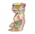
Circulatory SystemDescription - Carnegie Stage 22 embryo (27mm Human embryo, approximate day 56) Considerable remodeling takes place to transform the 3 components of the early circulation into a system that resembles the adult circulation, albeit with some important differences. As the embryo receives its oxygenated blood from the umbilical veins, its manner of distribution of the blood differs from the adult. However, after birth the infant must rapidly change to the adult pattern. Thus, the embryonic circulation is similar to the adult circulation, however the embryo’ vasculature features a number of shunts that allow the blood to bypass certain areas. In the late embryonic/foetal circulation, oxygenated blood from the placental travels through the umbilical vein, where it is shunted to the inferior vena cava, via a bypass called the ductus venosus. In doing so, the blood mixes with some portal blood and deoxygenated blood from the legs and the trunk. The blood enters the right atrium, where most of it is shunted to the left atrium via an open communication between the two – the foramen ovale. Although the superior vena cava delivers deoxygenated blood from the head and neck to the right atrium, the two streams of blood mix minimally. From the left atrium, the oxygenated blood is forced into the aorta, traveling first to the head and neck, and then supplying the trunk and legs. The blood then returns to the placenta, via the umbilical arteries. The vasculature of the head is formed primarily by remodeling of the arch arteries. The area above the heart – the aortic sac – forms a pair of horns, with the right horn forming the brachiocephalic artery and the left horn forming the common carotid artery. Arch arteries 1 and 2 regress, with the first forming part of the maxillary artery. The 3rd artery contributes to the internal carotid arteries, while the 4th is incorporated into the aortic arch and the 6th contributes to the ductus venosus. Blood supply to the limbs is established via an axial artery that develops along the central axis of the limb bud and eventually joins with the intersegmental arteries from the aorta. In the upper limb, the axial artery forms the brachial artery; in contrast, the axial artery of the lower limb is a continuation of the internal iliac artery. It mostly degenerates, with blood supply established by the external iliac artery. Between weeks 5 and 8, the early heart tube undergoes considerable remodeling, creating the four-chambered structure of the adult heart. Septation events split the four chambers of the heart, and simultaneous looping of the heart tube brings the atria and ventricles into correct anatomical position. This also brings the superior and inferior vena cavae into the right atrium, while the left atrium experiences outgrowth of the pulmonary vein. This branches twice to form four pulmonary veins that grow towards the lungs. |
|
|
|
|
|
|
These 3d movies were part of the UNSW Medical degree Independent Learning Project (ILP) prepared by Aashish Kumar (2006).
Glossary Links: A | B | C | D | E | F | G | H | I | J | K | L | M | N | O | P | Q | R | S | T | U | V | W | X | Y | Z | Numbers | Symbols | Movies
Cite this page: Hill, M.A. (2026, Mayıs 5) Embryology Cardiovascular 3D stage 22 Movie. Retrieved from https://embryology.med.unsw.edu.au/embryology/index.php/Cardiovascular_3D_stage_22_Movie
- © Dr Mark Hill 2026, UNSW Embryology ISBN: 978 0 7334 2609 4 - UNSW CRICOS Provider Code No. 00098G