Book - Vertebrate Zoology (1928) 33
Vertebrate Zoology G. R. De Beer (1928)
| Historic Disclaimer - information about historic embryology pages |
|---|
| Pages where the terms "Historic" (textbooks, papers, people, recommendations) appear on this site, and sections within pages where this disclaimer appears, indicate that the content and scientific understanding are specific to the time of publication. This means that while some scientific descriptions are still accurate, the terminology and interpretation of the developmental mechanisms reflect the understanding at the time of original publication and those of the preceding periods, these terms, interpretations and recommendations may not reflect our current scientific understanding. (More? Embryology History | Historic Embryology Papers) |
Chapter XXXIII The Ductless Glands
The ductless glands, or endocrine organs, are a group of structures remarkable no less for their function than for their mode of development, and their evolutionary history. The method of pouring out a secretion into the blood-stream instead of leading it away by a duct, is secondary, and some glands which are now ductless doubtless possessed ducts at earlier stages in evolution. Others, comprising the majority of the endocrine organs, were originally not glands at all, but structures which have become useless in the sense that their original function is not or cannot any longer be performed. They have become modified and their functions have changed in a remarkable manner. It is perhaps not without significance that so many of the ductless glands should have such a his- tory of structural and functional transformation. Another peculiarity which applies to several at least of these organs is that in development they arise from two separate rudiments, distinct in manner and place of origin, and even in the germ- layer from which they are formed.
The method of secreting into the blood-stream carries with it a property which cannot be possessed by glands secreting by means of definite ducts, for the latter can only communicate with definite and restricted spaces in the body, and the effects of such secretions must be only local. On the other hand, the blood circulates all over the body, carrying the endocrine secretions with it. These can therefore affect the body as a whole, and they are of immense importance both during development and during adult life in effecting correlations of the various parts with one another. The ductless glands act as a chemical mechanism of integration relying on the trans- portation of the stimulus (the secretions) through the vascular system; and this mechanism is complementary to that of nervous correlation and integration which involves not transportation of stimuli but conduction of impulses arising from stimuli along special paths, the nerves. " Secretin,' ' which is produced by the lining of the intestine and stimulates the pancreas to secrete, has been mentioned in Chapter XXVI.
The Thyroid
The thyroid was originally a longitudinal tract of ciliated and mucous-producing cells on the floor of the pharynx, called the endostyle. The endostyle is typically represented in Amphioxus (and in the Ascidians), where it is correlated with the ciliary method of feeding, and serves to make a moving " fly paper," on to which particles of food adhere and get carried safely back into the intestine (along the hyperpharyngeal groove), instead of getting carried out through the gill-slits by the outgoing current of water and lost. Such an endostyle is also present in the Ammoccete larva of Petromyzon. In the adult, however, it becomes closed off from the pharynx and sunk beneath it, and it gives rise to the vesicles of the thyroid. In all Gnathostomes the thyroid arises in development from the floor of the pharynx, and in some Selachii its cells still show traces of flagella. In the bony fish, the thyroid is not enclosed in a capsule of connective tissue, with the result that when it undergoes abnormal growth (goitre) it may become carcinomatous and give rise to a malignant cancer which invades the neighbouring tissues, including the bones. In the higher forms the thyroid is enclosed in a capsule.
The secretion of the thyroid increases the speed of the processes of metabolism in the body, and it has been said that it stands in the same relation to the body as the draught does to the fire. It plays an important part in the metamorphosis of amphibia, by promoting the growth of the (previously invisibly determined) regions into the organs which distinguish the tadpole from the adult frog or newt.
The Pituitary
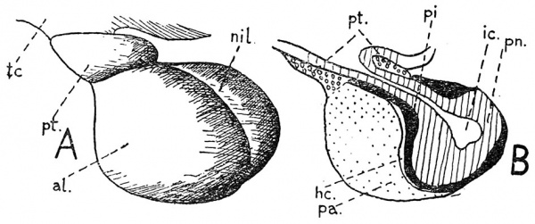
In all Craniates, the pituitary body is a composite organ formed from the hypophysis which grows in from the superficial ectoderm of the front of the head, and the infundibulum which is a down-growth from the floor of the forebrain. In Myxine these two constituents remain separated by connective tissue, but in all the remaining animals they are intimately connected and fused. In the Tetrapods it is possible to distinguish four parts in the pituitary, of which three (the anterior, intermedia, and tuberalis) arise from the hypophysis, and one (the nervosa) arises from the infundibulum. The intermedia is always (except in Myxine) plastered on to the nervosa, and the two together form the neuro-intermediate lobe. This is separated from an anterior lobe (formed of the anterior part) by the hypophysial cleft which represents the original cavity of the hypophysial ingrowth, Rathke's pocket.
Fig. 174. The pituitary body of a cat, seen, A, from the left side ; B, in longitudinal section. al, anterior lobe ; he, hypophysial cleft ; ic, infundibular cavity ; nil, neuro-intermediate lobe ; pa, pars anterior ; pi, pars intermedia ; pn, pars nervosa ; pt, pars tuberalis ; tc, floor of the brain.
In some animals, the hypophysial cleft becomes obliterated in the adult.
In evolution, the hypophysis appeared before the infundi- bulum, for in Amphioxus the latter is not represented, whereas the hypophysis is present in the form of the preoral pit. The preoral pit communicates with the (left) anterior head-cavity just as the hypophysis communicates with the premandibular somite in a number of Craniates. In the adult Amphioxus the preoral pit becomes absorbed in the oral hood and gives rise to the ciliated organ which produces a current of water towards the mouth. Thereafter it probably sank into the tissues and became a gland secreting by a duct into the mouth. This duct (which represents the open mouth of the cavity of Rathke's pocket) is preserved in Polyp terus, and Cyclostomes. In the latter, however, the duct has given rise to the large hypophysial sac which extends beneath the brain and has lost contact with the pituitary body. At the next stage in its evolution it must be imagined that the gland entered into relations with the infundibulum of the brain, and that it adopted the method of secreting into the blood-stream.
The functions of the pituitary are many, and they are only very imperfectly known. It must suffice to say that among these functions are those of : promotion of growth, control of blood-pressure, causing contractions of the uterus, expanding the black pigment-cells in the skin of amphibia, and stimulating the mammary glands to secrete milk.
The Adrenal
Like the pituitary, the adrenal bodies of the Tetrapods are composite structures. They are made up of an external cortex derived from the (mesodermal) coelomic epithelium, and a central medulla (chromafrlne tissue, so-called from its staining reactions) derived from the (ectodermal) cells which have migrated out from the nerve-tube in connexion with the sympathetic nerve-cells. In the fish, these two components are quite separate. The cortex of the adrenal is in them represented by the inter-renal, which, as its name implies, is situated between the kidneys. The medulla is represented by a number of supra-renal bodies which lie on or near the sympathetic nerve-chains, on each side of the aorta ; they are roughly segmental in arrangement. In the Cyclo- stomes, the supra-renals are closely associated with the ganglia of the dorsal roots, but the inter- renals are not well known.
Coming to the Tetrapods, the inter-renals and supra-renals are fused together to form the adrenal bodies, but in the more primitive forms such as the newts, these still resemble the fish in that they are not compact but form separate strips extending along the sympathetic nerve-chains, from the kidney to the anterior region of the thorax. The carotid gland, which is situated at the joint of the internal and external carotid arteries, is one of these.
The secretion of the medullary portion of the adrenal (adrenalin) has been synthetically prepared, but in spite of this fact, little is known of the functions of the gland, except that it produces effects similar to those due to stimulation through the sympathetic autonomic nervous system.
The Thymus
The thymus first appears in the fish as a series of paired upgrowths from the roof of the gill-slits. In the Selachians it is more or less segmental in its arrangement, but in higher forms the correspondence is lost, and the number of slits which contribute to it is reduced. It controls the formation of the shell, shell-membranes, and albumen in birds' eggs.
The Parathyroid
The name parathyroid is given to bodies which are usually situated close to or even in the thyroid, but which differ from the latter in their structure and method of development. They arise from the ventral regions of the 3rd and 4th visceral pouches in the Tetrapods, and are apparently absent in the fish.
The Pineal
The pineal eye has already been described in connexion with the sense organs. In the higher vertebrates this structure degenerates and is transformed into a gland.
The Pancreas
In addition to its function of producing enzymes for the purpose of digesting the food in the intestine, whither the enzymes are conducted by the pancreatic duct, the pancreas also functions as an organ of internal secretion. The tissue responsible for producing this internal secretion is that known as the islets of Langerhans, and its production is called insulin . The function of insulin is to store up glycogen in the liver, in which respect it is antagonised by the adrenalin. Diabetes is the result of faulty or non-functioning of the islets of Langerhans. In some Teleost fish, the endocrine islet- tissue may form little masses separate and apart from the ordinary pancreatic tissue, which secretes the digestive pan- creatic juice.
The " Puberty " Gland
The reproductive glands, ovary and testis, in the birds and mammals produce internal secretions which are concerned with the development and maintenance of the characters which distinguish one sex from the other. Since these secretions are essential for the proper sexual differentiation of the developing animals, the glands producing them have been called " puberty " glands.
The Corpus Luteum
The corpus luteum is the name given to what is really a temporary endocrine organ in the mammals. After an egg has vacated its Graafian follicle, the follicle undergoes changes resulting in the increase in size of the follicular cells, and the invasion of the follicle by connective tissue and blood-vessels. Should the egg liberated not get fertilised, the corpus luteum soon disappears. Should fertilisation result, however, and the blastocyst become attached to the wall of the uterus, the corpus luteum persists and increases in size, until the end of pregnancy. During this time it produces a secretion the functions of which are to prevent other eggs from being released from the ovary, and to control the growth of the uterus and the secretion of milk.
Literature
Riddle, O. Internal Secretions in Evolution and Reproduction. The Scientific Monthly, vol. 26, 1928. Swale Vincent. Internal Secretion and the Ductless Glands. Arnold, London, 1924.
| Historic Disclaimer - information about historic embryology pages |
|---|
| Pages where the terms "Historic" (textbooks, papers, people, recommendations) appear on this site, and sections within pages where this disclaimer appears, indicate that the content and scientific understanding are specific to the time of publication. This means that while some scientific descriptions are still accurate, the terminology and interpretation of the developmental mechanisms reflect the understanding at the time of original publication and those of the preceding periods, these terms, interpretations and recommendations may not reflect our current scientific understanding. (More? Embryology History | Historic Embryology Papers) |
- Vertebrate Zoology 1928: PART I 1. The Vertebrate Type as contrasted with the Invertebrate | 2. Amphioxus, a primitive Chordate | 3. Petromyzon, a Chordate with a skull, heart, and kidney | 4. Scyllium, a Chordate with jaws, stomach, and fins | 5. Gadus, a Chordate with bone | 6. Ceratodus, a Chordate with a lung | 7. Triton, a Chordate with 5-toed limbs | 8. Lacerta, a Chordate living entirely on land | 9. Columba, a Chordate with wings | 10. Lepus, a warm-blooded, viviparous Chordate PART II 11. The development of Amphioxus | 12. The development of Rana (the Frog) | 13. The development of Gallus (the Chick) | 14. The development of Lepus (the Rabbit) PART III 15. The Blastopore | 16. The Embryonic Membranes | 17. The Skin and its derivatives | 18. The Teeth | 19. The Coelom and Mesoderm | 20. The Skull | 21. The Vertebral Column, Ribs, and Sternum | 22. Fins and Limbs | 23. The Tail | 24. The Vascular System | 25. The Respiratory system | 26. The Alimentary system | 27. The Excretory and Reproductive systems | 28. The Head and Neck | 29. The functional divisions of the Nervous system | 30. The Brain and comparative Behaviour | 31. The Autonomic Nervous system | 32. The Sense-organs | 33. The Ductless glands | 34. Regulatory mechanisms | 35. Blood-relationships among the Chordates PART IV 36. The bearing of Physical and Climatic factors on Chordates | 37. The origin of Chordates, and their radiation as aquatic animals | 38. The evolution of the Amphibia : the first land-Chordates | 39. The evolution of the Reptiles | 40. The evolution of the Birds | 41. The evolution of the Mammalia | 42. The evolution of the Primates and Man | 43. Conclusions | Figures | Historic Embryology
Cite this page: Hill, M.A. (2024, April 23) Embryology Book - Vertebrate Zoology (1928) 33. Retrieved from https://embryology.med.unsw.edu.au/embryology/index.php/Book_-_Vertebrate_Zoology_(1928)_33
- © Dr Mark Hill 2024, UNSW Embryology ISBN: 978 0 7334 2609 4 - UNSW CRICOS Provider Code No. 00098G

